Optical Coherence Tomography in Dermatology
High-resolution imaging technology for noninvasive skin lesion diagnosis and disease monitoring.

What is Optical Coherence Tomography?
Optical coherence tomography (OCT) is a noninvasive diagnostic imaging method that provides real-time, high-resolution cross-sectional and en face images of the superficial layers of the skin in vivo. Using an infrared broadband light source, OCT allows clinicians to visualize skin architecture and detect pathological changes at depths of 1 to 2 mm with exceptional resolution ranging from 3 to 15 micrometers, depending on the specific system used.
The technology fills a critical gap in dermatological imaging by offering superior resolution compared to ultrasound while maintaining the noninvasive nature of clinical examination. Unlike traditional histopathological investigation, OCT preserves the natural morphology of skin structures without requiring tissue removal or processing.
How OCT Works: Technical Principles
OCT operates as an interferometric imaging method, typically implemented using fiber optics. The fundamental principle involves splitting light into two distinct beams: a probe beam and a reference beam. The probe beam focuses on the area of clinical interest within the skin, where it becomes backscattered by tissue structures. This backscattered light is then remixed with the reference beam.
When both beams match within the coherence length of the light source, interference occurs, creating the detailed images characteristic of OCT. The axial resolution—the ability to distinguish structures along the depth axis—depends on the bandwidth and coherence length of the infrared light source. The lateral resolution, which determines clarity in horizontal directions, is determined by the focusing objective of the imaging system.
Recent technical improvements have enabled OCT to achieve lateral resolution as fine as 3 to 7.5 micrometers. This advancement allows clinicians to visualize subtle architectural differences within skin layers, including the papillary dermis vasculature and hair follicles.
Polarisation-Sensitive OCT
Polarisation-sensitive OCT represents an advanced variant that provides additional contrast mechanisms for skin imaging. This technology measures the time delay between returning light consisting of two orthogonally polarised states. Light traveling through highly aligned fibres, such as organized collagen structures, accumulates phase-retardance between these polarized states, resulting in a visible banding pattern known as birefringence. In contrast, light passing through regions of disorganized or damaged fibres shows no such banding pattern, allowing discrimination of tissue organization status.
Clinical Applications in Skin Cancer Diagnosis
Basal Cell Carcinoma Detection
OCT has been most extensively studied for diagnosing basal cell carcinoma (BCC), the most common form of human malignancy. The technology offers attractive potential for noninvasive detection of early-stage disease. In a comprehensive analysis of 142 OCT images evaluated without additional clinical information, experienced dermatologists achieved diagnostic accuracy with sensitivity ranging from 86 to 95% and specificity from 81 to 98% for BCC identification.
Nonmelanoma Skin Cancers
OCT enables comprehensive evaluation of nonmelanoma skin cancers, including squamous cell carcinoma (SCC). The imaging characteristics of these lesions display distinctive patterns that allow differentiation from normal skin and benign lesions. Decreased pixel intensities in SCC images likely reflect the formation of cellular aggregates, small blood vessels, and deposition of fibrous collagen within the epidermis.
Melanoma and Pigmented Lesions
Melanoma and benign pigmented lesions present characteristic OCT appearances due to melanin content within the tissue. Pigmented lesions demonstrate irregular scattering patterns that distinguish them from unpigmented structures. OCT imaging provides quantitative analysis capabilities through pixel intensity profiles, allowing differentiation between melanomas and benign pigmented lesions such as seborrheic keratosis.
Actinic Keratosis Evaluation
Actinic keratosis lesions display distinctive OCT features including hyperkeratosis and apparent thickening of the entire epidermis. Despite this epidermal enlargement, the demarcation between the epidermis and dermis remains detectable on OCT images, facilitating accurate diagnosis and monitoring of lesion progression.
Role in Inflammatory and Connective Tissue Disorders
OCT holds considerable promise for diagnosing and monitoring inflammatory skin diseases. These conditions frequently present with abnormalities of cutaneous vasculature, and pathology is typically localized to the upper regions of the skin, making them ideal targets for OCT assessment.
Psoriasis and Eczema
Both psoriasis and atopic eczema demonstrate characteristic OCT findings including thickening of the stratum corneum and epidermis. Inflammatory infiltration and dermal edema result in decreased scattering intensity, while dilated blood vessels become readily apparent. The quantification and monitoring capabilities of OCT allow clinicians to assess therapeutic efficacy over time without requiring repetitive skin biopsies.
Contact Dermatitis Assessment
Early studies of OCT in inflammatory dermatology evaluated imaging in contact dermatitis, demonstrating the technology’s ability to visualize inflammatory changes in this common condition.
Darier’s Disease
In Darier’s disease, OCT can detect acantholytic papules and visualize the disruption of normal epidermal architecture associated with this genetic disorder.
Specialized Dermatological Applications
Nail Unit Examination
The nail unit can be effectively investigated using OCT. Healthy nail plates appear as well-demarcated structures presenting mostly with signal-rich parallel layers, occasionally displaying a granular pattern. Differences in scattering caused by different structural components and variations in layer thickness allow quantification of pathological changes including acanthosis, atrophy, or edema within the nail unit.
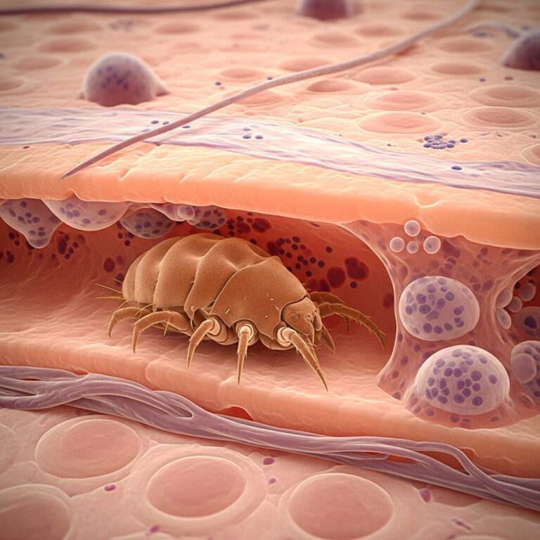
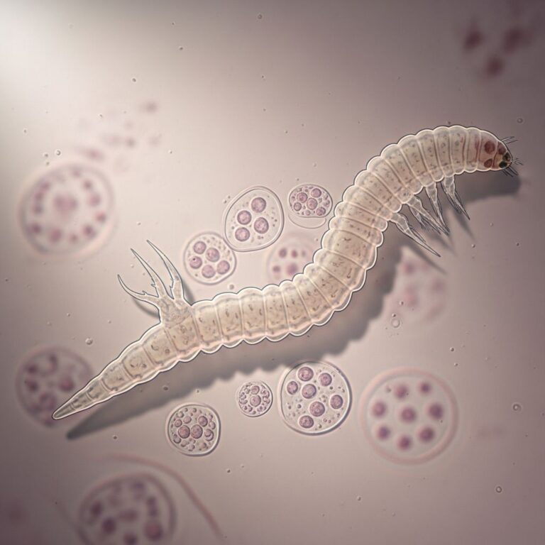
Parasitic Infestation Visualization
OCT enables visualization of parasitic infestations, providing diagnostic information about the depth and extent of parasitic invasion within the skin.
Additional Indications
Beyond skin cancers and inflammatory diseases, OCT finds application in evaluating:
- Bullous diseases and their subclinical manifestations
- Tattoos and tattooing complications
- Hemangiomas and vascular lesions
- Diabetic foot ulcers for depth assessment and healing monitoring
- Hypertrophic scars and burn wound assessment
- Systemic sclerosis with collagen content analysis
Advantages Over Traditional Diagnostic Methods
OCT provides several significant advantages compared to conventional dermatological imaging and diagnostic approaches:
- Noninvasive nature: Unlike histopathology, OCT visualizes unaltered tissue morphology without tissue removal or processing
- Real-time imaging: High-speed data acquisition enables immediate visualization of skin structures
- Quantification capability: Allows objective measurement of epidermal thickness, dermal changes, and collagen content
- Therapeutic monitoring: Eliminates need for repetitive biopsies when assessing treatment response in skin cancers and inflammatory diseases
- Complementary imaging: Works synergistically with dermoscopy, high-frequency ultrasound, and confocal laser scanning microscopy
Limitations and Clinical Challenges
Despite significant potential, adoption of OCT within dermatology has progressed slowly. The primary limitation stems from the high degree of optical scattering within skin tissue, which impedes deeper penetration of infrared light. This optical scattering characteristic makes it difficult for optical imaging to achieve the depth penetration achieved in other tissue types.
OCT angiography (OCTA), a functional extension developed for visualizing blood vessels, typically cannot be effectively used in dermatological applications due to the high scattering within the epidermis and dermis before reaching deeper vascular layers.
Technical Advances and Future Directions
Researchers continue advancing OCT technology for dermatological applications. The Department of Biomedical Engineering at Duke University has pioneered developments including:
- Introduction of new sources of optical contrast
- Incorporation of spectroscopic information for enhanced burn depth assessment
- Integration of Raman spectroscopy for detection of biological analytes
- Methods for increasing depth penetration beyond conventional limits
Dual-axis OCT represents an emerging approach utilizing distinct illumination and detection apertures to triangulate regions of interest up to 2.5 mm beneath the skin surface, extending imaging depth beyond conventional single-axis systems. Additionally, the selection of wavelength, detection approaches, advanced beam geometries, and sophisticated analysis techniques can further improve diagnostic capabilities for assessing tissue structures at various depths.
Clinical Integration and Practical Considerations
The high dynamic range exceeding 100 dB achieved by modern OCT systems allows for penetration depths of 1 mm or more, depending on tissue properties and wavelength selection. These characteristics position OCT as a valuable addition to the dermatologist’s diagnostic armamentarium.
OCT usage is particularly reasonable when noninvasive therapies are applied, as it avoids repetitive biopsies while enabling control of therapeutic efficacy. In actinic keratosis and squamous cell carcinoma management, OCT can provide estimation of infiltrative growth patterns crucial for treatment planning.
Comparing OCT with Other Diagnostic Modalities
| Imaging Modality | Depth Penetration | Resolution | Invasiveness | Key Advantages |
|---|---|---|---|---|
| OCT | 1-2 mm | 3-15 μm | Noninvasive | Real-time, quantifiable, structures preserved |
| Dermoscopy | Surface only | Lower | Noninvasive | Cost-effective, portable, rapid assessment |
| Confocal Laser Microscopy | Up to 250 μm | 0.5-1 μm | Noninvasive | Cellular resolution, excellent detail |
| High-Frequency Ultrasound | 5+ mm | Higher | Noninvasive | Greater depth penetration |
| Histopathology | N/A | Cellular | Invasive | Gold standard diagnostic reference |
Frequently Asked Questions
Q: How deep can OCT imaging penetrate into skin?
A: OCT systems typically achieve penetration depths of 1 to 2 millimeters, with advanced dual-axis systems extending to approximately 2.5 millimeters. This depth allows visualization of the entire epidermis and substantial portions of the dermis.
Q: What is the resolution of OCT compared to other imaging methods?
A: OCT provides lateral resolution between 3 and 15 micrometers depending on the system. This resolution is superior to ultrasound but lower than confocal laser microscopy, positioning OCT as a complementary imaging technique.
Q: Can OCT replace skin biopsy for cancer diagnosis?
A: While OCT achieves high diagnostic accuracy for basal cell carcinoma (86-95% sensitivity, 81-98% specificity), it is best used as a complementary diagnostic tool rather than a complete replacement for histopathological examination in all cases.
Q: Is OCT painful or uncomfortable for patients?
A: No, OCT is completely noninvasive and painless. Patients experience no tissue trauma, discomfort, or adverse effects during imaging.
Q: What are the main clinical applications of OCT in dermatology?
A: Primary applications include diagnosis of skin cancers (particularly basal cell carcinoma), evaluation of inflammatory diseases (psoriasis, eczema), monitoring treatment response, assessment of nail disorders, and evaluation of other skin conditions including parasitic infestations and vascular lesions.
Q: Why has OCT adoption been slow in dermatology despite its advantages?
A: The high degree of optical scattering within skin tissue limits penetration depth and image quality compared to other tissue types. This technical limitation has restricted clinical adoption, though ongoing research continues improving performance.
Q: How does polarisation-sensitive OCT differ from standard OCT?
A: Polarisation-sensitive OCT measures time delay between orthogonally polarised light states, revealing tissue organization through birefringence patterns. Organized collagen shows characteristic banding, while disorganized tissue lacks this pattern, providing additional diagnostic contrast.
References
- Optical coherence tomography in dermatology — National Center for Biotechnology Information (PubMed). 2013. https://pubmed.ncbi.nlm.nih.gov/23314617/
- Optical coherence tomography in dermatology — SPIE Digital Library. 2013. https://www.spiedigitallibrary.org/journals/journal-of-biomedical-optics/volume-18/issue-6/061224/Optical-coherence-tomography-in-dermatology/10.1117/1.JBO.18.6.061224.full
- Applications and future directions for optical coherence tomography in dermatology — Oxford Academic (British Journal of Dermatology). 2001. https://academic.oup.com/bjd/article/184/6/1014/6697827
- OCT – Polarisation-sensitive OCT — Sheffield Dermatology Research. Accessed January 2026. https://sheffielddermatologyresearch.com/oct
- Use of Optical Coherence Tomography images to Differentiate Between Normal Skin, Skin Lesions and Melanoma: A Pilot Study — Clinical and Medical Images. Accessed January 2026. https://clinandmedimages.org/use-of-optical-coherence-tomography-images-to-differentiate-between-normal-skin-skin-lesions-and-melanoma-a-pilot-study/
- Optical Coherence Tomography Noninvasively Images Skin Structure — Photonics Magazine. Accessed January 2026. https://www.photonics.com/Articles/Optical-Coherence-Tomography-Noninvasively-Images/a70374
- In Vivo High-Definition Optical Coherence Tomography — JAMA Dermatology. Accessed January 2026. https://jamanetwork.com/journals/jamadermatology/fullarticle/1918743
Similar Articles


Read full bio of medha deb