Orf Virus: Clinical Presentation and Management
Understanding orf virus lesions, progression stages, and evidence-based treatment approaches for healthcare providers.

Orf Virus: Clinical Presentation and Management Overview
Orf virus, scientifically known as Orfviridae, is a contagious viral infection that primarily affects sheep and goats but can transmit to humans through direct contact with infected animals. The disease is also known as contagious ecthyma, contagious pustular dermatitis, or scabby mouth in animals, and farmyard pox in humans. This zoonotic pathogen has gained clinical significance due to its ability to cause painful lesions and potential complications in both animal and human populations. Understanding the clinical presentation, progression, and management of orf virus infection is essential for veterinarians, healthcare providers, and individuals working with livestock.
Clinical Presentation in Animals
In animals, particularly young sheep and goats, orf virus causes characteristic pustular lesions that typically begin on the lips, muzzle, and mouth areas. The virus represents the natural host range for these species, and infected animals develop painful vesicular lesions that can impede normal feeding and care activities. Secondary locations affected by the virus include teats, vulva, scrotum, ears, and coronary bands of the hooves. The pain associated with these lesions can lead to severe complications including anorexia, abandonment of offspring, and lameness in affected animals.
Animal lesions are characterized by the formation of crusts and scabs around the nose, mouth, teats, and other affected areas, which is why the condition is commonly referred to as scabby mouth. While orf virus infections are typically self-limiting in animals, secondary bacterial infections can occur and complicate the clinical course.
Clinical Presentation in Humans
Humans typically contract orf virus through direct contact with infected animals or contaminated fomites, a process known as a host-switch zoonotic transmission. Unlike the oral involvement seen in animals, human infections usually present on the dorsal surfaces of the hands, fingers, and forearms at areas of contact with infected animals. The initial presentation involves the development of painful lesions that can be infectious, potentially spreading to other areas of the body if scratched or touched.
Associated systemic symptoms may accompany the local lesions, including mild fever, tiredness, and swelling of lymph nodes in the neck and underarm areas. Red streaks along the lymph channels (lymphangitis) with enlargement of lymph glands on the inner side of the elbow and under the arm (lymphadenopathy) are not uncommon presentations. In some cases, patients may develop secondary bacterial infections that can complicate the clinical picture and prolong healing.
Lesion Progression Stages
The progression of orf virus lesions follows a characteristic sequence of morphological changes spanning approximately four to six weeks in immunocompetent individuals. Understanding these stages is crucial for accurate diagnosis and patient counseling regarding expected disease progression.
Stage 1: Maculopapular Stage
The infection begins with the maculopapular stage, characterized by the development of erythematous macules and papules at the site of contact with infected material. After an incubation period of approximately five to six days, small papules emerge that gradually become ulcerative in nature. These initial lesions are typically painful and may cause localized discomfort.
Stage 2: Target Stage
As the infection progresses, lesions evolve into targetoid nodules surrounded by a red halo with a necrotic center, characteristic of the target stage. This distinctive appearance may resemble other dermatological conditions, making clinical differentiation important for accurate diagnosis.
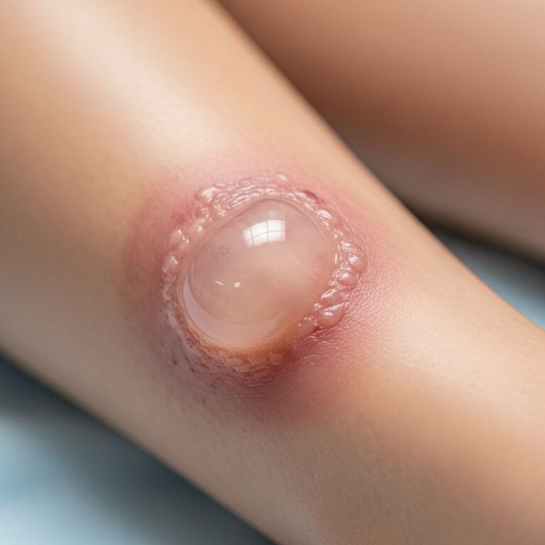
Stage 3: Acute Stage
During the acute stage, nodules begin to weep and may become increasingly uncomfortable. This stage represents the peak of inflammation and represents the most infectious phase of the lesions, as viral particles are actively shed from the weeping surfaces.
Stage 4: Regenerative Stage
The regenerative stage is marked by the drying of the nodule, with the beginning of the healing process. The lesion begins to transition toward resolution with the formation of initial crusting.
Stage 5: Papilloma Stage
The nodule develops a dry crust and becomes papillomatous, with the formation of characteristic tiny black spots on the lesion surface. This stage represents continued progression toward healing with progressive crusting and reduced viral shedding.
Stage 6: Regression Stage
In the final regression stage, the lesion begins to regress and eventually resolves completely. Most lesions heal without scarring in immunocompetent individuals, though the timeline can extend beyond six weeks in some cases.
Associated Dermatological Manifestations
Beyond the primary orf lesions, various secondary dermatological manifestations can develop. Erythema multiforme, a secondary rash associated with the presence of orf virus, may develop 10–14 days after the onset of orf and results in targetoid macules, plaques, and blisters on the hands, feet, face, arms, and legs. Less distinctive red rashes, termed toxic erythemas, also occur in some patients.
Rarely, blistering disorders such as bullous pemphigoid can develop, particularly in immunocompromised individuals. These complications are more common in patients with compromised immune function, including those with HIV infection or undergoing chemotherapy.
Pathological Features and Diagnosis
Histopathological examination of orf lesions reveals distinctive features that aid in diagnosis. The virus demonstrates eosinophilic intracytoplasmic inclusions and ballooning degeneration characteristic of poxvirus infections. Ballooning reticular degeneration due to cellular edema is another common finding, though the stratum corneum is usually intact.
Dermal changes include heavy lymphohistiocytic infiltrative changes, and staining procedures such as methenamine silver, Ziehl-Neelsen (acid-fast), and periodic acid-Schiff are typically negative, aiding in differentiation from other conditions. Spongiform degeneration in follicular structures and polymorphic infiltrate may be present in the lower dermis.
Early diagnosis is crucial to prevent disease worsening and to implement appropriate management strategies. The combination of clinical presentation, histopathological findings, and patient history of exposure to infected animals typically supports diagnosis.
Treatment and Management Strategies
Orf viral infection demonstrates a favorable prognosis in immunocompetent individuals, with lesions being self-limiting. Symptoms typically resolve spontaneously within a 4- to 6-week period without specific antiviral therapy, and most cases require nothing more than reassurance and expectant care.
Supportive Care
For immunocompetent patients, supportive and at-home care constitutes the primary management approach. Pain management, hygiene maintenance, and prevention of secondary bacterial infection are key components of supportive care. Patients should be advised to keep lesions clean and dry, and to avoid scratching to prevent autoinoculation to other body areas.
Antiviral Therapy
While no widely approved medical treatments exist for orf virus infection, several antiviral approaches have demonstrated efficacy, particularly in immunocompromised patients. Topical cidofovir, a potent blocker of the novel viral form of DNA-dependent RNA polymerase utilized by Orfviridae, has achieved nearly ubiquitous use as first-line antiviral therapy. Cidofovir has demonstrated activity against multiple orthopox and parapox viruses, with animal models revealing its effectiveness in reducing orf lesion development in lambs and rescuing mice with orf lesions.
Brincidofovir represents another antiviral agent that has been studied in ecthyma contagiosum and other orthopoxviruses with demonstrated effectiveness, including in vitro studies using rabbits. Systemic interferon-α has been employed to treat immunocompromised patients with persistent or severe lesions. Additionally, antiviral agents that inhibit nucleoside metabolism have been proposed as effective treatments.
Mechanical Eradication
Mechanical approaches to lesion management may be implemented alongside or instead of antiviral therapy. Cryotherapy and imiquimod application represent mechanical eradication methods that can be used if needed. However, surgical debridement should be avoided as this can prolong recovery.
Management in Immunocompromised Patients
In immunocompromised hosts, orf can grow to several centimeters, and the resultant morphology may mimic tumors and pyogenic granuloma, which may require further management. These patients benefit from more aggressive therapeutic approaches, including antiviral therapy and close monitoring for complications. Treatment decisions in immunocompromised patients should be individualized based on lesion severity, extent, and patient immune status.
Prevention and Vaccination
Vaccination has proven particularly effective for orf virus prevention due to the highly conserved and complex antigenicity of the pathogen. Many domesticated sheep and goat populations are routinely vaccinated with varying levels of success. While human vaccines are less commonly used, vaccination strategies have demonstrated efficacy in reducing disease incidence in animal populations.
In humans, recurrence of orf is rare, but when it does occur, subsequent episodes demonstrate diminished severity. This suggests that primary infection confers partial protective immunity against future infections.
Veterinary Considerations
In veterinary settings, management of orf in livestock requires careful attention to secondary infections and pain control. Procaine penicillin injections have demonstrated beneficial effects in severe cases of orf in sheep. Treatment of secondary bacterial infections, particularly those involving Staphylococcus aureus, is important as these can cause severe dermatitis.
For foot infections related to orf virus, topical treatment with oxytetracycline spray is typically effective. Management of fly strike through application of appropriate topical agents is vital, particularly during summer months when orf outbreaks are more likely to attract secondary fly infestation. Caregivers should always use protective equipment, including rubber gloves, when handling affected animals to minimize transmission risk.
Transmission Prevention and Infection Control
Given the contagious nature of orf virus, prevention of transmission is an important consideration. The virus spreads through direct contact with infected animals or through fomites contaminated with viral particles. Healthcare providers and individuals working with livestock should maintain strict hygiene practices, including the consistent use of protective gloves when handling potentially infected animals or patients.
The rash associated with orf is infectious and can spread to other areas of a person’s body if they scratch or touch the sores. Educating patients about avoiding contact with their own lesions and maintaining good hand hygiene is essential for preventing autoinoculation and limiting disease spread.
Prognosis and Expected Outcomes
The prognosis for orf virus infection is generally favorable in immunocompetent individuals. The disease typically resolves completely within four to six weeks without intervention, and lesions generally heal without scarring. Most patients experience only localized morbidity during the active infection phase, with systemic symptoms being mild and self-limited.
In immunocompromised patients, the prognosis is more variable. These individuals may experience prolonged disease courses, larger lesions, and increased risk of complications. However, with appropriate management including antiviral therapy, even severely immunocompromised patients can achieve lesion resolution, though the timeline may be extended.
Frequently Asked Questions
Q: How long does orf virus infection typically last?
A: In immunocompetent individuals, orf virus infection typically resolves within 4 to 6 weeks without specific treatment. The lesions progress through six characteristic stages during this period, ultimately healing without scarring in most cases.
Q: Can orf virus cause serious complications?
A: While complications are rare in immunocompetent individuals, immunocompromised patients may develop larger lesions, bacterial superinfection, or secondary conditions such as erythema multiforme or bullous pemphigoid. These complications typically resolve with appropriate management.
Q: Is there a cure for orf virus infection?
A: Orf virus infection is self-limiting, meaning it resolves on its own without specific cure. Most immunocompetent patients require only supportive care. For immunocompromised patients, antiviral agents such as cidofovir may help accelerate healing.
Q: How is orf virus transmitted to humans?
A: Humans contract orf virus through direct contact with infected animals, particularly sheep and goats, or through contact with contaminated materials (fomites) from infected animals. The virus enters through breaks in the skin at sites of contact.
Q: Can you catch orf virus from another person?
A: While the primary source of infection is infected animals, the orf rash itself is infectious and can spread to other areas of an infected person’s body through scratching or touching. However, person-to-person transmission is uncommon compared to animal-to-human transmission.
Q: What should I do if I suspect orf virus infection?
A: If you suspect orf virus infection, consult a healthcare provider for diagnosis, particularly if you work with livestock or have direct animal contact. Early diagnosis can help guide appropriate management and prevent disease worsening. Maintain good hygiene and avoid touching or scratching lesions to prevent spread.
References
- Contagious Ecthyma (Orf Virus Infection) — Montana Department of Livestock, Animal Health Bureau. 2013-12. https://liv.mt.gov/_docs/Animal-Health/Newsletters/2013_12_Dec_MOH.pdf
- Orf Viral Infection — StatPearls, National Center for Biotechnology Information (NCBI). 2025. https://www.ncbi.nlm.nih.gov/books/NBK562191/
- Diagnosis and Treatment of Orf — Vet Times, Division of Veterinary Clinical Sciences, University of Edinburgh. 2024. https://www.vettimes.com/news/vets/livestock/diagnosis-andtreatment-of-orf
- Orf — DermNet, New Zealand Dermatological Society. 2024. https://dermnetnz.org/topics/orf
- About Orf Virus (Sore Mouth Disease) — Centers for Disease Control and Prevention (CDC). 2025. https://www.cdc.gov/orf-virus/about/index.html
- What Is Orf Virus? — WebMD Medical Reference. 2025. https://www.webmd.com/a-to-z-guides/what-is-orf-virus
- Orf on the Farm — University of Wisconsin Environment, Health & Safety. 2020-08. https://ehs.wisc.edu/wp-content/uploads/sites/1408/2020/08/EHS-ARS-GUI-032-V01.pdf
Similar Articles


Read full bio of Sneha Tete