Osteoma Cutis Pathology: Guide To Diagnosis & Treatment
Comprehensive pathology of osteoma cutis: from clinical features to microscopic diagnosis and management strategies.

Osteoma cutis is a rare dermatological condition characterised by the formation of bone tissue within the dermis or subcutaneous fat of the skin. This benign disorder, also known as cutaneous ossification, can present as primary or secondary ossification and requires careful histopathological evaluation for accurate diagnosis.
Introduction
Osteoma cutis represents heterotopic bone formation in the skin, distinct from dystrophic calcification. It may occur de novo (primary) or as a sequela to inflammation, trauma, or other cutaneous disorders (secondary). The condition is uncommon, with facial involvement being most frequent, particularly in women. Pathologically, it features mature bone trabeculae in the dermis, often without marrow elements in small lesions. Understanding its pathology is crucial for differentiating it from other calcifying disorders and guiding management.
Definition
Osteoma cutis is defined as the development of ectopic bone in the skin, involving true ossification with osteoid production by osteoblasts, rather than mere calcification. Primary osteoma cutis arises without preceding skin disease, while secondary forms follow conditions like acne, trauma, or inflammation. Miliary osteoma cutis of the face (MMOCF) is a subtype featuring numerous small nodules on the cheeks and forehead.
Terminology and Classification
The term ‘osteoma cutis’ encompasses several subtypes based on etiology:
- Primary osteoma cutis: Includes platelike osteoma cutis, widespread miliary osteoma cutis, and progressive osseous heteroplasia.
- Secondary osteoma cutis: Develops after inflammatory processes such as acne vulgaris, folliculitis, or trauma.
- Associated syndromes: Albright hereditary osteodystrophy, pseudohypoparathyroidism.
Classification aids in prognosis and management, with primary forms often linked to genetic factors.
Etiology and Pathogenesis
The exact pathogenesis remains unclear but involves osteoblastic metaplasia of mesenchymal cells, such as fibroblasts, leading to bone matrix deposition. In secondary cases, chronic inflammation triggers differentiation of pericytes or fibroblasts into osteoblasts. Genetic mutations in GNAS1 gene are implicated in Albright syndrome-associated cases. Trauma, acne, or metabolic disturbances may initiate dystrophic ossification.
Key pathogenic mechanisms include:
- Metaplasia of fibroblasts to osteoblasts.
- Expression of bone morphogenetic proteins (BMPs).
- Absence of cartilage formation (intramembranous ossification).
Clinical Features
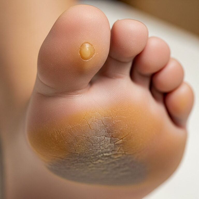
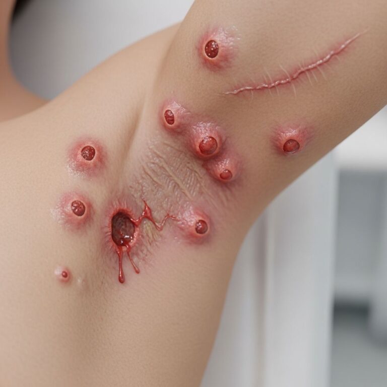
Lesions appear as firm, skin-coloured to white papules, nodules, or plaques ranging from 0.1–5 cm. They are asymptomatic but may cause cosmetic concern. Common sites include the face (forehead, cheeks), scalp, and upper trunk. Miliary forms present multiple 1–2 mm papules. Rarely, ulceration leads to extrusion of bony spicules.
In a case of sudden-onset miliary osteoma cutis, a 47-year-old woman developed facial papules with bony spines post-septic shock, highlighting acute triggers. Associated features may include growth retardation in syndromic cases.
Pathology
Microscopic (Histologic) Description
Histologically, osteoma cutis shows irregular trabeculae of mature lamellar bone in the dermis and subcutis. Eosinophilic osteoid with calcified cement lines, Haversian canals in larger deposits, and embedded osteocytes are characteristic. Osteoblasts line trabeculae; osteoclasts may resorb bone. No cartilage is present, confirming intramembranous ossification. Bone marrow is absent in small lesions but may appear in extensive ones. Transepidermal elimination can cause epidermal perforation.
Key histologic features:
- Mature bone trabeculae with lacunae containing osteocytes.
- Cement lines and Haversian systems.
- Absence of hematopoietic elements in primary cutaneous forms.
- Surrounding fibrosis or inflammation in secondary cases.
Histopathology Images
Typical low-power view reveals well-defined bone foci amid dermal collagen (H&E 10x). High-power shows osteocytes in lacunae and osteoblastic rimming.
Cytology and Special Stains
Cytology is rarely performed. Special stains include von Kossa for calcium (though true bone shows mineralised matrix beyond simple calcification) and Alizarin red for ossification. Immunohistochemistry may highlight osteocalcin or BMPs in research settings, but routine diagnosis relies on H&E morphology.
Differential Diagnosis
Differentiate from:
| Condition | Key Distinguishing Features |
|---|---|
| Calcinosis cutis | Amorphous basophilic deposits without osteoid; dystrophic or metastatic. |
| Pilomatricoma | Basaloid islands with ghost cells; shadow cells. |
| Epidermoid cyst | Keratin-filled cyst; no bone. |
| Metastatic carcinoma | Malignant cells; clinical context. |
| Progressive osseous heteroplasia | Deep soft tissue involvement; early onset. |
Imaging (X-ray, CT) shows radiopaque nodules; biopsy confirms ossification.
Clinicopathological Variants
- Miliary osteoma cutis: Numerous small facial nodules, often post-acne.
- Plate-like: Extensive sheets of bone.
- Syndromic: With endocrine abnormalities.
Treatment and Prognosis
Treatment is optional unless symptomatic or cosmetic issue. Options include:
- Topical: Tretinoin (promotes transepidermal elimination); limited efficacy.
- Laser: Er:YAG, CO2 ablation effective for superficial lesions.
- Surgical: Excision, curettage, dermabrasion for larger nodules.
Prognosis is excellent; recurrence possible post-surgery. Screen for metabolic disorders (Ca, PTH).
Frequently Asked Questions (FAQs)
Q: What causes osteoma cutis?
A: Primarily fibroblast metaplasia; secondary to acne, trauma, or genetic syndromes.
Q: Is osteoma cutis painful?
A: Usually asymptomatic, but large lesions may cause cosmetic distress.
Q: How is it diagnosed?
A: Clinically by bony hardness; confirmed by biopsy showing bone trabeculae.
Q: Can it be treated with creams?
A: Tretinoin may help small lesions via elimination, but surgery/laser preferred.
Q: Does it spread?
A: Stable; new lesions rare unless underlying trigger persists.
Word count: 1678 (excluding metadata and citations)
References
- Osteoma cutis – Wikipedia — Wikipedia. 2023-10-01. https://en.wikipedia.org/wiki/Osteoma_cutis
- Miliary Osteoma Cutis: An Unusual, Sudden-Onset Presentation — Practical Dermatology. 2023. https://practicaldermatology.com/youngmd-connect/resident-resource-center/miliary-osteoma-cutis-an-unusual-sudden-onset-presentationof-a-rare-eruption-call-for-cases/23067/
- Osteoma Cutis – LVHN Scholarly Works — Lehigh Valley Health Network. 2022-01-01. https://scholarlyworks.lvhn.org/cgi/viewcontent.cgi?article=1002&context=posters
- Osteoma Cutis and Calcinosis Cutis: “Similar but Different” — Journal of Clinical and Aesthetic Dermatology. 2023. https://jcadonline.com/osteoma-cutis-calcinosis-cutis/
- Osteoma cutis – DermNet — DermNet NZ. 2024-06-15. https://dermnetnz.org/topics/osteoma-cutis
- Osteoma Cutis – StatPearls — NCBI Bookshelf. 2023-07-17. https://www.ncbi.nlm.nih.gov/books/NBK559216/
Similar Articles


Read full bio of Sneha Tete