Pericardial Effusion: Symptoms, Causes & Treatment
Understanding pericardial effusion: causes, symptoms, complications, and treatment options for fluid around the heart.

Pericardial effusion is a medical condition characterized by an abnormal buildup of fluid in the space around your heart. This fluid accumulates in the pericardium, which is the protective sac that surrounds your heart. While the pericardium normally contains a small amount of fluid to cushion and lubricate the heart, pericardial effusion occurs when excess fluid collects in this space. Understanding this condition is essential for recognizing symptoms and seeking appropriate medical care, as pericardial effusion can range from mild to life-threatening depending on various factors.
What Is Pericardial Effusion?
The pericardium is a two-layered membrane that encloses the heart and the roots of the major blood vessels. In a healthy state, the pericardium contains only a small amount of lubricating fluid—typically less than 50 milliliters—that allows the heart to move smoothly within the sac as it contracts and expands. This natural fluid serves as a protective barrier and reduces friction between the heart and surrounding tissues.
Pericardial effusion develops when this delicate balance is disrupted and excess fluid accumulates in the pericardial space. Depending on the volume and rate of fluid accumulation, this condition can have varying degrees of severity. When fluid builds up slowly over weeks or months, the pericardium has time to stretch and accommodate the additional volume. However, when fluid accumulates rapidly, the pericardium cannot expand quickly enough, leading to increased pressure on the heart and potentially dangerous complications.
Causes of Pericardial Effusion
Pericardial effusion can develop for numerous reasons, and identifying the underlying cause is crucial for determining the appropriate treatment approach. The causes are diverse and can range from inflammatory conditions to traumatic injuries.
Common Causes
Infections represent one of the most frequent causes of pericardial effusion. Viral infections are particularly common, including those caused by influenza, COVID-19, and other respiratory viruses. Bacterial infections, though less common, can also lead to pericardial effusion and may be more severe. Tuberculosis remains an important cause in certain geographic regions and populations.
Inflammatory and autoimmune conditions frequently contribute to pericardial effusion development. These include lupus, rheumatoid arthritis, and other connective tissue disorders that can trigger inflammation in the pericardium. Heart and circulatory problems such as heart attack, heart failure, and high blood pressure can also result in fluid accumulation around the heart.
Malignancy represents a significant cause of pericardial effusion, particularly in advanced cancer cases where tumors can metastasize to the pericardium or obstruct lymphatic drainage. Additionally, kidney disease and hypothyroidism can lead to pericardial effusion through various mechanisms affecting fluid balance and metabolism.
Other Contributory Factors
Injuries and trauma to the chest can cause immediate or delayed pericardial effusion as blood or fluid accumulates in the pericardial space. Radiation therapy to the chest, commonly used in cancer treatment, may damage the pericardium and lead to effusion months or even years after treatment. Certain medications and medical procedures, including cardiac surgery or catheterization, can also trigger pericardial effusion as a complication.
In many cases, the cause of pericardial effusion remains unknown, a condition referred to as idiopathic pericardial effusion. Interestingly, research indicates that pericardial effusions comprise approximately 70% of pericardial disease cases seen in clinical settings, with roughly 19.5% of these developing into cardiac tamponade if left untreated.
Symptoms of Pericardial Effusion
The symptoms of pericardial effusion vary significantly depending on the amount of fluid present, the speed of accumulation, and whether complications like cardiac tamponade have developed. Many patients with mild effusions may experience no symptoms at all, while others may present with various clinical manifestations.
Early Warning Signs
When pericardial effusion does cause symptoms, chest pain is often the most prominent complaint. This pain typically originates behind the breastbone and may radiate to the shoulder or neck. The pain often worsens when lying flat or taking deep breaths, and may improve when sitting upright or leaning forward. Shortness of breath is another common symptom, resulting from the pressure the effusion places on the lungs and airways.
Palpitations—the sensation of a rapid, fluttering, or irregular heartbeat—frequently accompany pericardial effusion as the heart attempts to compensate for reduced pumping efficiency. Patients may also experience fatigue and a general sense of weakness, as organs receive decreased blood flow due to the heart’s compromised pumping ability.
Additional Manifestations
When a pericardial effusion is large enough to exert pressure on surrounding tissues or nerves, additional symptoms may develop. These can include a persistent cough, particularly in cases where the effusion presses on the airways or esophagus. Some patients report experiencing syncope or fainting episodes, especially if the effusion causes subclinical cardiac tamponade with elevated intrapericardial pressure affecting cardiac output during activities that increase intrathoracic pressure.
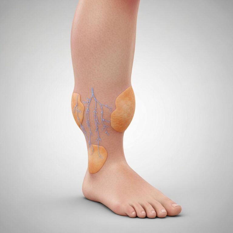
Swelling in the legs and ankles may occur as fluid accumulates and venous pressure increases. Abdominal bloating and fullness are also possible, reflecting systemic congestion resulting from inadequate cardiac output.
Complications: Understanding Cardiac Tamponade
While pericardial effusion itself can be managed in many cases, the most serious potential complication is cardiac tamponade. This life-threatening condition occurs when fluid pressure in the pericardium becomes so great that it prevents the heart’s chambers from expanding and filling with blood properly.
How Cardiac Tamponade Develops
Cardiac tamponade represents a medical emergency that can be rapidly fatal without immediate intervention. As pericardial fluid accumulates and pressure increases, the right atrium and right ventricle—the heart’s chambers that receive blood returning from the body—become compressed. This compression prevents these chambers from filling adequately, which reduces the amount of blood available for the heart to pump to the rest of the body.
The severity and speed of cardiac tamponade development depend critically on how quickly the fluid accumulates. When effusion develops slowly, the pericardium has time to stretch and expand, allowing it to accommodate relatively large volumes of fluid before symptoms become critical. Conversely, rapid fluid accumulation can produce cardiac tamponade with smaller fluid volumes because the pericardium cannot stretch quickly enough to accommodate the fluid without significantly increasing pressure.
Symptoms of Cardiac Tamponade
Classic signs of cardiac tamponade include the “Beck’s triad”: elevated jugular venous pressure (distended neck veins), muffled heart sounds on examination, and hypotension (low blood pressure). Patients typically present with severe dyspnea, anxiety, altered mental status, and signs of shock including cool, clammy skin. The diagnosis may be suspected based on hemodynamic findings including a narrow pulse pressure and elevated central venous pressure.
Without immediate treatment, cardiac tamponade leads to cardiogenic shock—a state of acute heart failure where the heart cannot pump enough blood to maintain organ perfusion. Cardiogenic shock is ultimately fatal without rapid intervention. Recognition of tamponade is therefore critical, as emergency pericardial drainage can be life-saving.
Other Potential Complications
Beyond cardiac tamponade, pericardial effusion can lead to other serious complications. Recurrent effusions may develop in some patients, requiring repeated drainage procedures or more definitive treatment. In cases of malignant pericardial effusion or those associated with HIV/AIDS, prognosis tends to be poor, with increased risk of complications and mortality.
A rare but serious complication is pericardial decompression syndrome, which can occur following drainage of large effusions. This syndrome results when fluid is removed too rapidly, allowing the collapsed heart to suddenly reexpand and potentially leading to acute pulmonary edema and hemodynamic instability.
Diagnosis and Imaging
Accurate diagnosis of pericardial effusion relies on a combination of clinical evaluation and advanced imaging techniques. Healthcare providers employ multiple diagnostic modalities to assess the presence, size, and hemodynamic significance of pericardial fluid.
Echocardiography (ultrasound of the heart) is the primary imaging modality for detecting and assessing pericardial effusion. Transthoracic echocardiography provides clear visualization of fluid around the heart and can evaluate whether the effusion is causing hemodynamic compromise. This technique also helps determine whether fluid is loculated (compartmentalized) or free-flowing.
Additional imaging modalities complement echocardiography in specific clinical scenarios. Cardiac magnetic resonance imaging (MRI) excels at visualizing pericardial inflammation and confirming the diagnosis of constrictive pericarditis. Computed tomography (CT) imaging effectively demonstrates pericardial calcification, which may be present in chronic constrictive pericarditis. Chest X-rays may show an enlarged cardiac silhouette when significant effusion is present, though this finding is nonspecific.
Treatment Options
Treatment of pericardial effusion depends on multiple factors including the underlying cause, the amount of fluid present, the rate of accumulation, and whether the effusion is causing hemodynamic compromise or symptoms.
Conservative Management
Mild pericardial effusions that develop slowly and cause no symptoms may not require immediate treatment. In such cases, close monitoring with serial echocardiography is appropriate to ensure the effusion is not enlarging. Treatment of the underlying cause—such as managing kidney disease, correcting hypothyroidism, or treating infection—may allow gradual reabsorption of the fluid.
Pericardiocentesis
For larger or symptomatic effusions, pericardiocentesis is often the first-line therapeutic intervention. This procedure involves inserting a needle or catheter into the pericardial space under imaging guidance (typically echocardiographic guidance) to drain excess fluid. Pericardiocentesis can be both diagnostic—allowing analysis of the fluid to identify underlying causes—and therapeutic, providing immediate relief of symptoms and prevention of cardiac tamponade.
The volume and characteristics of drained fluid provide valuable diagnostic information. Laboratory analysis may reveal evidence of infection, malignancy, or other specific conditions guiding further management. In emergency situations with cardiac tamponade, pericardiocentesis can be rapidly life-saving.
Surgical Interventions
For effusions that recur after pericardiocentesis or those associated with constrictive pericarditis, surgical approaches become necessary. Pericardiectomy—surgical removal of the pericardium—represents the definitive surgical treatment for recurrent effusions and constrictive pericarditis. “Radical pericardiectomy,” meaning removal of all pericardium or as much as absolutely possible, offers the best outcomes for preventing recurrent symptoms. Incomplete removal of pericardium can result in recurrent symptoms requiring additional surgery.
Pericardial window procedures, which create an opening in the pericardium to allow continuous drainage into the pleural space, may be employed in selected cases, particularly for malignant effusions. These approaches allow fluid to drain naturally while avoiding repeated procedures.
Medical Management
For effusions related to inflammatory conditions like acute pericarditis, non-steroidal anti-inflammatory drugs (NSAIDs) and colchicine may reduce inflammation and promote fluid reabsorption. Treatment of underlying conditions—such as antibiotics for infectious pericarditis, diuretics and afterload reduction for heart failure-related effusion, or cancer therapy for malignant effusions—addresses the root cause and may facilitate resolution.
Prognosis and Outlook
The prognosis of pericardial effusion varies considerably depending on the underlying etiology, the amount of fluid, and whether complications have developed. Effusions occurring for unknown reasons (idiopathic) generally carry a good prognosis, with many resolving spontaneously or responding well to medical management.
Conversely, effusions related to malignancy, trauma, or HIV/AIDS tend to have poorer prognoses with higher complication rates and greater treatment difficulty. Survival rates are particularly concerning for patients whose pericardial effusions are linked to cancer or HIV/AIDS, making early diagnosis and aggressive management critical in these populations.
With appropriate treatment at specialized pericardial disease centers, outcomes can be optimized. Referral to tertiary centers with expertise in pericardial disease management is particularly important for complex cases requiring surgery, as these centers have significantly better operative outcomes than general hospitals.
When to Seek Medical Attention
Patients experiencing chest pain, shortness of breath, palpitations, or syncope should seek medical evaluation promptly. Sudden onset of these symptoms warrants emergency medical attention, as they may indicate cardiac tamponade. Individuals with known pericardial effusion require regular follow-up with their healthcare provider to monitor for changes in effusion size or development of complications.
Frequently Asked Questions
Q: What is the difference between pericarditis and pericardial effusion?
A: Pericarditis is inflammation of the pericardium and often precedes pericardial effusion. While pericarditis refers to the inflammatory process, pericardial effusion specifically refers to the accumulation of fluid as a result of that or other conditions. Pericardial effusions only present in about 50% of patients with pericarditis.
Q: Can pericardial effusion resolve on its own?
A: Yes, small or mild effusions may resolve spontaneously with treatment of the underlying condition. However, large effusions typically require intervention and will not resolve without fluid removal or definitive treatment of the underlying cause.
Q: Is pericardial effusion always dangerous?
A: Not all pericardial effusions are immediately dangerous. Mild effusions may cause no symptoms and require only monitoring. The danger depends on the amount of fluid, how quickly it accumulates, and whether it progresses to cardiac tamponade.
Q: How quickly can pericardial effusion become life-threatening?
A: Rapidly accumulating effusions can become life-threatening within hours, while slowly developing effusions may take weeks or months to cause critical problems. Rapid accumulation of even modest volumes can cause cardiac tamponade and cardiogenic shock, which can be fatal within minutes to hours without treatment.
Q: What is pericardial decompression syndrome?
A: This is a rare but serious complication occurring when fluid is drained too quickly from a large pericardial effusion. The sudden reexpansion of the heart can cause acute pulmonary edema and hemodynamic instability. Healthcare providers prevent this by draining effusions more slowly when necessary.
Q: Where should pericardial effusion surgery be performed?
A: Complex pericardial effusion cases requiring surgery should be managed at specialized tertiary pericardial disease centers. These centers perform significantly more procedures annually (50-100 cases yearly) compared to average hospitals, resulting in substantially better operative outcomes.
References
- Abstract 3908: Pericardial Disease: Uncommon or Under-diagnosed? — American Heart Association, Circulation. 2009. https://www.ahajournals.org/doi/10.1161/circ.120.suppl_18.S884-d
- Pericardial Effusion: Symptoms & Causes — Cleveland Clinic. 2024. https://my.clevelandclinic.org/health/diseases/17351-pericardial-effusion
- Recurrent Pericardial Effusions and Pericarditis Due to Yellow Nail Syndrome — National Center for Biotechnology Information, PMC. 2023. https://pmc.ncbi.nlm.nih.gov/articles/PMC10107038/
- Don’t Judge a Book by Its Cover: Unusual Presentations of Pericardial Disease — Cleveland Clinic Journal of Medicine. 2024. https://www.ccjm.org/content/92/2/109
- Avoiding Misdiagnosis and Undertreatment in Pericarditis — Cleveland Clinic Consult QD. 2024. https://consultqd.clevelandclinic.org/resource-for-avoiding-misdiagnosis-and-undertreatment-in-pericarditis
- American College of Cardiology Concise Clinical Guidance on Pericarditis — Cleveland Clinic Consult QD. 2024. https://my.clevelandclinic.org/podcasts/cardiac-consult/american-college-of-cardiology-concise-clinical-guidance-on-pericarditis
Similar Articles


Read full bio of medha deb