Periocular Dermatitis: Causes, Symptoms & Treatment
Understanding periocular dermatitis: symptoms, diagnosis, and effective treatment options.

Periocular Dermatitis: Causes, Symptoms, and Treatment
Periocular dermatitis, also known as periorbital dermatitis, is a common inflammatory skin condition characterized by small, red, scaly papules and pustules that develop around the eye and eyelid area. This condition affects the delicate skin surrounding the eyes and can cause significant discomfort and cosmetic concern for affected individuals. Understanding the nature of periocular dermatitis, its underlying causes, and available treatment options is essential for effective management and symptom resolution.
Definition and Overview
Periocular dermatitis represents a form of inflammatory dermatosis localized to the orbital and periorbital regions. The condition is distinguished by its characteristic appearance and distribution pattern around the eyes. Unlike other facial dermatologic conditions, periocular dermatitis specifically affects the thin, sensitive skin of the eyelids and surrounding areas, making it both aesthetically noticeable and potentially uncomfortable for patients.
Causes and Etiological Factors
Periocular dermatitis can result from multiple causative factors. The two primary categories of contact-related triggers include allergic contact dermatitis (ACD) and irritant contact dermatitis (ICD). These distinct mechanisms require different diagnostic and management approaches.
Allergic Contact Dermatitis represents an immune-mediated response triggered by exposure to specific allergens. Common allergens affecting the periocular region include components found in mascara, eyeshadow, eyeliner, contact lens solutions, eye drops, and eyeglass frames, particularly those containing nickel. The condition develops when the immune system becomes sensitized to an offending substance and subsequently reacts upon re-exposure.
Irritant Contact Dermatitis results from direct chemical damage to the skin barrier without requiring prior sensitization. Irritants commonly encountered in the periocular area include preservatives in cosmetics and eye care products, fragrances, and harsh skincare ingredients. This type of dermatitis typically develops more rapidly following exposure compared to allergic reactions.
Beyond contact-related causes, topical steroid use can precipitate or exacerbate periocular dermatitis, particularly when steroids are applied chronically to the delicate periocular skin. Other potential contributing factors include seborrheic dermatitis, rosacea variants, and infectious agents.
Clinical Presentation and Symptoms
Periocular dermatitis typically presents with several characteristic symptoms affecting the eye region:
- Redness (erythema) surrounding the eye and eyelid
- Scaling and flaking of the skin
- Swelling (edema) of the eyelids and periorbital tissues
- Itching and pruritus
- Burning sensation, particularly in irritant contact dermatitis cases
- Small raised bumps (papules) and fluid-filled lesions (vesicles)
The distribution pattern and characteristics of these symptoms can provide diagnostic clues regarding the underlying etiology. For instance, nickel allergy from eyeglasses may result in erythema concentrated near the cheek and eyebrow while sparing the eyelids, whereas reactions to mascara components typically localize to only the eyelid area. Patients with irritant contact dermatitis frequently report a “burning” sensation, while those with allergic contact dermatitis more commonly experience itching.
In cases of prolonged irritation, clinical presentation may progress beyond simple erythema and papules to include exudation, crusting, and in chronic situations, lichenification—a thickening and hardening of the skin.
Diagnosis and Diagnostic Methods
Clinical Examination
Diagnosis of periocular dermatitis is primarily established through clinical evaluation and detailed patient history. The characteristic appearance of small, red, scaly papules and pustules around the eye region is usually readily recognizable to experienced clinicians. The general morphology and distribution pattern of lesions often permit preliminary diagnosis based on visual examination alone.
Differential Diagnosis
Distinguishing periocular dermatitis from other skin conditions is crucial for appropriate management. Key differential diagnoses include:
- Acne: Distinguished from periocular dermatitis by the presence of comedones (blackheads and whiteheads)
- Rosacea: Characterized by inflammatory papules and pustules primarily affecting the central face, with associated telangiectatic erythema and flushing
- Seborrheic dermatitis: Presents with different distribution and clinical characteristics
- Contact dermatitis from other body regions: Must be differentiated to identify the specific offending agent
Additional Diagnostic Testing
While clinical diagnosis is typically sufficient, additional diagnostic modalities may be employed when initial evaluation is inconclusive. Skin biopsy, though generally not necessary for routine cases, may be considered in atypical presentations or when treatment response is inadequate. Biopsy findings typically demonstrate non-specific inflammation with perifollicular or perivascular lymphohistiocytic infiltration.
Patch testing and the repeated open application test (ROAT) prove valuable in determining the specific causative allergen, particularly when allergic contact dermatitis is suspected. These tests involve controlled exposure to potential allergens to identify which substances trigger an immune response in the individual patient.
If a bacterial component is suspected, bacterial culture may be obtained. Potassium hydroxide (KOH) preparation and scraping can be performed if Candida species are considered contributory to the clinical presentation. In severe cases, diascopy performed by an experienced clinician may provide additional diagnostic information.
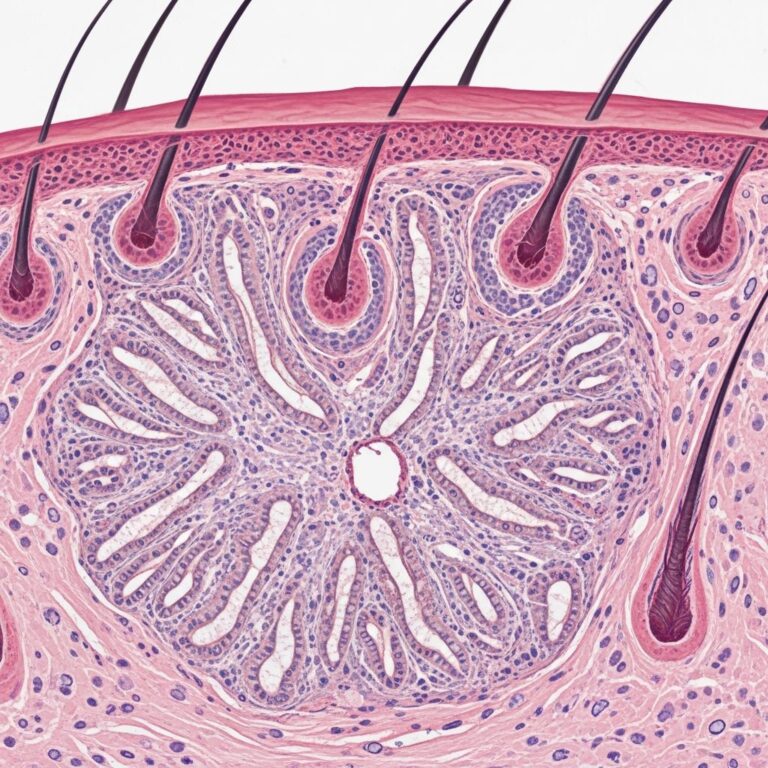
Pathology and Histopathology
Histological examination of periocular dermatitis specimens reveals non-specific inflammation characterized by perifollicular or perivascular lymphohistiocytic infiltration affecting vellus hair follicles. These microscopic findings reflect the underlying immune-mediated inflammatory process but are generally not specific enough to identify the causative agent.
Treatment and Management Strategies
General Treatment Principles
Management of periocular dermatitis revolves around identifying and avoiding triggering agents combined with symptomatic relief through topical medications. A critical first step involves discontinuing all periocular use of cosmetics and skin care products, regardless of whether a specific offending agent has been identified. Patients should be counseled to wash the face with warm water only during the acute phase.
For individuals with suspected allergic contact dermatitis, patch testing with potential allergens can facilitate identification of triggering substances. A practical home test involves applying new products to the neck or forearm for 5-7 days before facial application to detect potential reactions.
Topical Therapeutic Options
Topical therapy proves effective for mild disease and may be used as monotherapy or in combination with oral medications for moderate to severe cases.
Topical Antibiotics: First-line topical treatments include metronidazole cream or gel, clindamycin lotion or gel, and erythromycin gel. These antibiotics are particularly effective due to their potent anti-inflammatory properties beyond their antimicrobial action. Topical preparations demonstrate good penetration into the periocular tissues while minimizing systemic absorption.
Topical Calcineurin Inhibitors: Tacrolimus ointment and pimecrolimus cream represent effective alternatives, particularly for periocular seborrheic dermatitis. These medications modulate immune function and reduce inflammation through distinct mechanisms compared to corticosteroids. Calcineurin inhibitors offer the advantage of avoiding steroid-related side effects and can be used for extended durations without the concern for skin atrophy or systemic absorption complications.
Topical Sulfur Preparations and Azelaic Acid: Sulfacetamide-sulfur combinations and azelaic acid gel provide additional topical options with anti-inflammatory and antimicrobial properties.
Topical Corticosteroids: While topical corticosteroids may be employed for brief periods in mild cases, caution is warranted regarding their chronic use in the periocular region. Long-term corticosteroid application can precipitate complications including skin atrophy, steroid sensitization, and adrenal suppression. Furthermore, chronic topical steroid use can paradoxically trigger or perpetuate periocular dermatitis through steroid-induced flares.
Oral Therapeutic Options
Oral antibiotics are employed for moderate to severe disease or when topical therapies prove insufficient. Tetracycline-class antibiotics including tetracycline, doxycycline, and minocycline represent first-line oral options. These medications provide dual benefit through antimicrobial and anti-inflammatory mechanisms. Treatment duration typically spans 6 to 12 weeks, with gradual tapering as clinical improvement occurs.
Treatment Response and Healing Phases
Periocular dermatitis typically responds well to appropriate treatment, though resolution requires patience and adherence to management strategies. The healing process typically progresses through distinct phases:
Initial Regression Phase: During early treatment, inflammation begins subsiding with gradual reduction in redness and papule prominence. Patients may experience dryness and peeling as the inflamed tissue resolves. During this phase, continued use of gentle skincare products is essential while harsh exfoliants must be avoided.
Progressive Improvement: As therapy continues, skin quality progressively normalizes with continued reduction in inflammatory signs and symptoms. Treatment may be required for up to three months in some cases.
Management of Steroid-Induced Flares
A significant consideration in periocular dermatitis management involves recognizing and appropriately handling steroid-induced exacerbations. When periocular dermatitis is triggered by topical steroid application, the primary treatment recommendation involves discontinuing steroid use. However, abrupt cessation can precipitate rebound flaring; therefore, gradual tapering under medical supervision proves necessary. This nuanced approach prevents iatrogenic harm while allowing safe transition away from steroids.
When to Consult a Dermatologist
Professional dermatologic evaluation is warranted in several clinical scenarios:
- Symptoms persist despite discontinuing steroid use and implementing gentle skincare routines
- Rash or bumps extend beyond the initial affected area
- Significant discomfort, burning, or itching develops
- Recurrent flare-ups demonstrate poor response to conservative management
- Diagnostic uncertainty exists regarding the underlying condition
Because periocular dermatitis can mimic other conditions including acne, rosacea, contact dermatitis, and seborrheic dermatitis, accurate professional diagnosis forms the foundation for effective treatment.
Frequently Asked Questions
What causes periocular dermatitis?
Periocular dermatitis primarily results from allergic or irritant contact dermatitis triggered by cosmetics, eye care products, eyeglass frames, or topical steroids. Less commonly, it may result from seborrheic dermatitis, rosacea variants, or infections.
How is periocular dermatitis diagnosed?
Diagnosis is typically clinical based on characteristic appearance and patient history. Patch testing can identify specific allergens. Skin biopsy is reserved for atypical presentations. Additional testing such as bacterial culture or KOH preparation may be performed when indicated by clinical suspicion.
What is the best treatment for periocular dermatitis?
Treatment involves identifying and avoiding triggering agents combined with topical medications. First-line options include topical antibiotics (metronidazole, clindamycin, erythromycin) or calcineurin inhibitors (tacrolimus, pimecrolimus). Oral antibiotics are used for moderate to severe cases. Topical corticosteroids should be used cautiously and briefly to avoid paradoxical worsening.
How long does periocular dermatitis take to resolve?
Treatment duration varies but typically requires 6 to 12 weeks. Mild cases may respond more quickly to topical therapy, while moderate to severe cases requiring oral antibiotics necessitate longer treatment courses with gradual medication tapering.
Can periocular dermatitis return?
Yes, periocular dermatitis can be persistent or recurrent. Long-term management strategies tailored to individual patients help prevent recurrence. Ongoing avoidance of identified triggers is essential for maintaining remission.
References
- Periorbital Dermatitis: Causes, Symptoms, and Treatment Options — Conlon Eye Institute. Accessed January 2026. https://conloneyeinstitute.com/periorbital-dermatitis-symptoms-treatment-options/
- Perioral Dermatitis – StatPearls — National Center for Biotechnology Information (NCBI). Updated January 2026. https://www.ncbi.nlm.nih.gov/books/NBK525968/
- Perioral Dermatitis – Dermatologic Disorders — Merck Manuals Professional Edition. January 2026. https://www.merckmanuals.com/professional/dermatologic-disorders/acne-and-related-disorders/perioral-dermatitis
- Periocular Dermatitis (Periorbital Dermatitis): Symptoms with Images — DermNet New Zealand. Accessed January 2026. https://dermnetnz.org/topics/periocular-dermatitis
- Signs Perioral Dermatitis is Healing: Key Phases Explained — US Dermatology Partners. January 2026. https://www.usdermatologypartners.com/blog/what-is-perioral-dermatitis/
- Perioral Dermatitis — MedlinePlus Medical Encyclopedia. National Library of Medicine. Updated January 2026. https://medlineplus.gov/ency/article/001455.htm
- Periocular (Periorbital) Dermatitis — EyeWiki. American Academy of Ophthalmology. January 2026. https://eyewiki.org/Periocular_(Periorbital)_Dermatitis
Similar Articles


Read full bio of Sneha Tete