Peripheral Vascular Disease: Causes, Symptoms, and Treatment
Comprehensive guide to understanding PVD: Learn about causes, symptoms, diagnosis, and effective treatment options.

Understanding Peripheral Vascular Disease
Peripheral vascular disease (PVD), also known as peripheral artery disease (PAD), is a circulatory condition characterized by the narrowing of blood vessels that carry blood to the extremities, most commonly the legs and feet. This chronic disease develops when fatty deposits accumulate in the arterial walls, restricting normal blood flow and oxygen delivery to affected tissues. PVD represents a significant manifestation of systemic atherosclerosis and affects millions of people worldwide, particularly those over 50 years of age.
When blood vessels become narrowed or partially blocked, the tissues they supply receive insufficient oxygen, leading to a range of symptoms that can impact quality of life and increase the risk of serious cardiovascular complications. Early recognition and appropriate management are essential to prevent disease progression and minimize the risk of severe outcomes such as tissue damage, infection, or amputation.
What Causes Peripheral Vascular Disease?
The primary cause of peripheral vascular disease is atherosclerosis, a progressive condition in which fatty plaques accumulate along the inner walls of arteries. This buildup narrows the vessel lumen, reducing the diameter available for blood flow and creating resistance that prevents adequate oxygen-rich blood from reaching peripheral tissues.
Risk Factors Contributing to PVD Development
Several modifiable and non-modifiable factors increase the risk of developing peripheral vascular disease:
Smoking is one of the most significant controllable risk factors. Tobacco use damages the endothelium (inner lining of blood vessels), promotes inflammation, and accelerates plaque formation.
Diabetes increases PVD risk substantially, as high blood sugar levels damage blood vessel walls and promote atherosclerotic changes. Diabetic patients often develop PVD earlier and experience more severe disease progression.
High blood pressure (hypertension) contributes to arterial wall damage and stiffness, facilitating plaque deposition and narrowing.
High cholesterol and elevated triglycerides provide the lipid building blocks for plaque formation. Low-density lipoprotein (LDL) cholesterol, often called “bad cholesterol,” is particularly atherogenic.
Age is a non-modifiable risk factor. The prevalence of PVD increases significantly in individuals over 65 years, though it can develop earlier in those with multiple risk factors.
Family history of cardiovascular disease indicates genetic predisposition to atherosclerosis and vascular disease.
Obesity and sedentary lifestyle contribute to metabolic dysfunction, increased inflammation, and reduced cardiovascular fitness, all promoting PVD development.
Kidney disease is associated with accelerated atherosclerosis and increased PVD risk.
Recognizing Symptoms of Peripheral Vascular Disease
Many individuals in the early stages of PVD experience no symptoms, which is why the condition is often discovered incidentally during evaluation for other health concerns. However, as arterial narrowing progresses, characteristic symptoms typically emerge.
Common Symptoms
Intermittent claudication is the hallmark symptom of PVD. This condition manifests as cramping, aching, tiredness, or heaviness in the leg muscles, typically during physical activity such as walking. The pain characteristically improves with rest, as the legs’ oxygen demand decreases. The location of claudication pain corresponds to the site of arterial narrowing—calf pain suggests narrowing in the femoral or popliteal arteries, while hip or buttock pain indicates aortoiliac disease.
Leg and foot pain may occur at rest or during sleep, indicating more advanced disease with severely compromised blood flow.
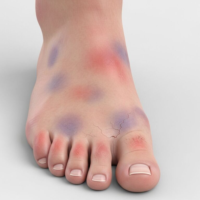
Coldness in the lower legs or feet, particularly when one extremity feels noticeably colder than the other, reflects reduced blood flow and heat delivery.
Color changes in the skin of the legs and feet may include paleness, a bluish or purplish discoloration (cyanosis), or a shiny appearance of the skin.
Non-healing wounds or ulcers on the toes, feet, or legs develop due to impaired blood supply and reduced oxygen availability for tissue repair.
Hair and nail changes include poor nail growth, decreased hair growth on the toes and legs, and thinning of skin.
Erectile dysfunction in males may indicate aortoiliac disease affecting blood flow to the pelvis.
Swelling (edema) in the legs or feet may occur, along with the development of varicose veins.
Gangrene, representing tissue death due to severe ischemia, is a serious complication requiring immediate medical intervention.
Diagnosis of Peripheral Vascular Disease
Accurate diagnosis requires a combination of clinical assessment, patient history, and specialized diagnostic testing.
Initial Evaluation
Your healthcare provider will perform a thorough physical examination, assessing pulses in your legs and feet, checking skin temperature and color, examining for wounds or ulcers, and evaluating muscle strength and sensation. A detailed history regarding symptoms, their onset, duration, and relationship to activity helps establish the diagnosis.
Diagnostic Tests
Ankle-brachial index (ABI) compares blood pressure in the ankle to that in the arm, providing an objective measure of lower extremity blood flow.
Duplex ultrasound uses sound waves to visualize blood vessels and measure blood flow velocity, identifying areas of narrowing.
Computed tomography (CT) angiography creates detailed images of blood vessels after contrast injection, showing the location and severity of narrowing.
Magnetic resonance angiography (MRA) provides similar vascular imaging without radiation exposure.
Coronary angiography involves direct catheterization and contrast injection into arteries, allowing visualization of even subtle narrowing and enabling simultaneous intervention if needed.
Complications of Untreated Peripheral Vascular Disease
Without appropriate management, PVD can progress to serious complications affecting not only the affected limb but systemic cardiovascular health.
Acute limb ischemia occurs when arterial blood flow suddenly decreases dramatically, causing severe pain and tissue damage if not rapidly treated.
Heart attack and stroke are significantly more likely in PVD patients, as atherosclerotic disease is typically systemic, affecting coronary and cerebral arteries as well.
Blood clots (thrombosis) may form in narrowed arteries, further compromising blood flow.
Tissue infection and gangrene develop when severely ischemic tissue becomes necrotic and infected, potentially requiring amputation.
Amputation becomes necessary in severe cases where limb salvage is impossible despite aggressive intervention.
Treatment Approaches for Peripheral Vascular Disease
Management of PVD aims to improve blood flow to affected tissues, relieve symptoms, and reduce the risk of systemic cardiovascular complications. Treatment typically progresses from conservative approaches to more invasive interventions based on symptom severity and response to initial therapy.
Lifestyle Modifications
Lifestyle changes form the foundation of PVD management and should be implemented regardless of other treatments pursued.
Smoking cessation is absolutely critical, as continued smoking accelerates disease progression and significantly increases complication risk.
Regular exercise, particularly supervised walking programs, improves symptoms by enhancing collateral blood vessel development and improving muscle efficiency. Structured exercise programs have been shown to increase walking distance and reduce claudication pain.
Healthy diet emphasizing vegetables, fruits, whole grains, and lean proteins while limiting saturated fats, trans fats, and sodium helps control weight, blood pressure, and cholesterol levels.
Weight management reduces cardiovascular risk factors and improves mobility.
Blood sugar control in diabetic patients prevents further vascular damage.
Pharmacological Treatment
Several medication classes are used to manage PVD:
Statins reduce cholesterol levels and help prevent further plaque accumulation while potentially stabilizing existing plaques.
Antihypertensive medications control blood pressure, protecting vessel walls from further damage.
Antiplatelet agents including aspirin and clopidogrel reduce blood clot risk by preventing platelets from aggregating.
Anticoagulants may be used when thrombotic risk is high.
Vasodilators relax smooth muscle in blood vessel walls, improving blood flow. Cilostazol is specifically used to improve walking distance in claudication.
Diabetes medications maintain tight glycemic control, preventing progressive vascular damage in diabetic patients.
Pain-relief medications help manage leg pain and improve activity tolerance.
Interventional and Surgical Procedures
When lifestyle modifications and medications fail to adequately manage symptoms or when disease is severe, minimally invasive or surgical interventions may be necessary.
Balloon angioplasty involves threading a catheter with an inflatable balloon to the narrowed artery segment. The balloon inflates to compress plaque and widen the arterial lumen, restoring blood flow.
Stent placement involves positioning a small mesh-like tube to maintain arterial patency after angioplasty, preventing restenosis.
Atherectomy uses specialized catheters to remove atherosclerotic plaque from vessel walls in selected cases.
Intravascular lithotripsy (IVL) uses shock waves to break up heavily calcified plaque, facilitating subsequent angioplasty.
Thrombolytic therapy dissolves acute blood clots by delivering clot-dissolving medications directly into affected arteries.
Bypass surgery creates alternative routes for blood flow around severely narrowed or blocked arteries using grafts from the patient’s own veins or synthetic materials.
Endarterectomy surgically removes plaque from arterial walls in select cases.
Frequently Asked Questions
Q: Is peripheral vascular disease curable?
A: There is no cure for PVD, but it can be effectively managed with lifestyle changes, medications, and interventional procedures to control symptoms and prevent progression.
Q: Can PVD affect my arms?
A: Yes, while PVD most commonly affects the legs, narrowed arteries can develop in the arms as well, though this is less frequent.
Q: How often should I exercise if I have PVD?
A: Supervised exercise programs typically recommend walking three to five times weekly for 30 to 60 minutes, gradually increasing distance and intensity as tolerated.
Q: What should I do if I develop a wound on my foot?
A: Seek immediate medical attention. In PVD patients, even minor wounds can become serious due to impaired healing. Proper wound care and infection prevention are critical.
Q: Can PVD lead to amputation?
A: Severe untreated PVD can progress to gangrene and tissue death, which may necessitate amputation. However, early detection and appropriate management can usually prevent this outcome.
References
- Peripheral Vascular Disease — HealthDirect (Australian Government Department of Health). 2024. https://www.healthdirect.gov.au/peripheral-vascular-disease
- Peripheral Artery Disease (PAD) – Symptoms and Causes — Mayo Clinic. 2024. https://www.mayoclinic.org/diseases-conditions/peripheral-artery-disease/symptoms-causes/syc-20350557
- Peripheral Artery Disease (PAD) – Diagnosis and Treatment — Mayo Clinic. 2024. https://www.mayoclinic.org/diseases-conditions/peripheral-artery-disease/diagnosis-treatment/drc-20350563
- Peripheral Vascular Disease: Diagnosis and Treatment — American Academy of Family Physicians. 2006. https://www.aafp.org/pubs/afp/issues/2006/0601/p1971.html
- Peripheral Arterial Disease (PAD) – Treatment — NHS (National Health Service). 2024. https://www.nhs.uk/conditions/peripheral-arterial-disease-pad/treatment/
- Peripheral Vascular Disease (PVD) — Yale Medicine. 2024. https://www.yalemedicine.org/conditions/peripheral-vascular-disease
- Peripheral Vascular Disease (PVD) Signs & Symptoms — Rush University Medical Center. 2024. https://www.rush.edu/conditions/peripheral-vascular-disease-pvd
Similar Articles


Read full bio of Sneha Tete