PET Scan: What It Is, Types, Purpose, Procedure & Results
Complete guide to PET scans: Understanding how positron emission tomography detects cancer, heart disease, and brain disorders.
What Is a PET Scan?
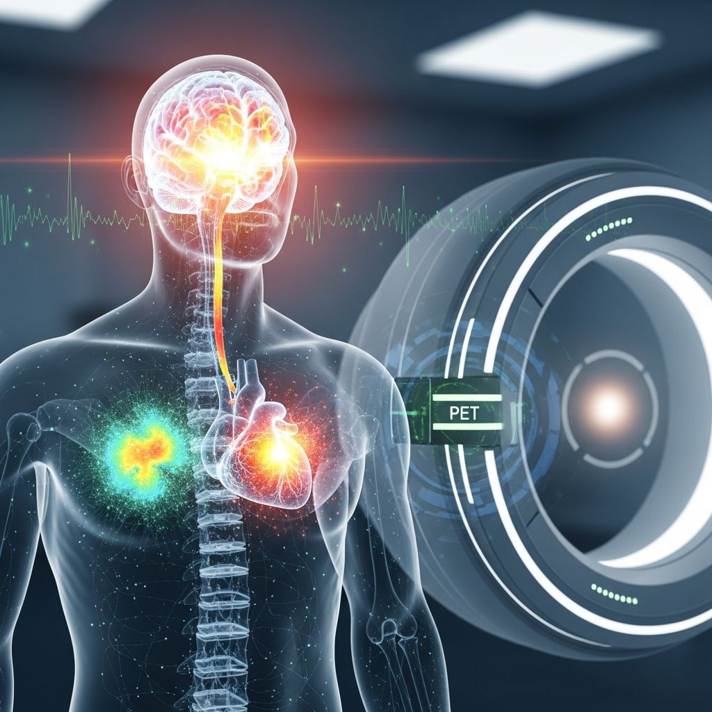
A positron emission tomography (PET) scan is a sophisticated medical imaging test that uses radioactive tracers to visualize how your body’s cells and organs are functioning. Unlike traditional imaging techniques such as computed tomography (CT) scans or magnetic resonance imaging (MRI) scans, which primarily show the structure of organs and tissues, PET scans focus on the biological activity and molecular processes occurring within your body. This unique capability allows healthcare providers to detect disease in its earliest stages, often before structural changes become visible on other imaging modalities.
During a PET scan, a healthcare provider injects a small amount of radioactive tracer into your bloodstream. This tracer travels through your body and accumulates in areas where cells are most active metabolically. Diseased cells, including cancer cells, typically absorb significantly more of the radiotracer than healthy cells do, creating what radiologists call “hot spots.” The PET scanner detects the radiation emitted by these tracers and produces detailed images that reveal the location and intensity of cellular activity.
How PET Scans Differ from Other Imaging Techniques
Understanding the differences between PET scans and other imaging methods helps clarify why your healthcare provider might recommend one test over another. Each imaging modality has distinct advantages and applications in modern medicine.
CT scans use X-ray technology to create detailed cross-sectional images of your body. They excel at showing the anatomical structure of organs and bones but provide limited information about how tissues are actually functioning.
MRI scans use powerful magnetic fields and radio waves to generate highly detailed images of soft tissues. While MRI provides exceptional anatomical detail without radiation exposure, it similarly captures structural rather than functional information.
PET scans operate on an entirely different principle. By measuring vital functions such as blood flow, oxygen use, and blood sugar (glucose) metabolism, PET scans reveal what your cells are actually doing at the molecular level. This functional perspective enables detection of abnormalities that may not yet be visible structurally.
Hybrid Imaging: PET/CT and PET/MRI Scanners
Modern medical imaging has evolved to combine the strengths of multiple technologies. A PET/CT scanner performs both a PET scan and a CT scan simultaneously using the same machine, producing three-dimensional images that merge functional information with anatomical detail. This combination allows for more accurate diagnosis and better localization of abnormalities.
Additionally, some hospitals now offer hybrid PET/MRI scanners, representing cutting-edge imaging technology. These systems create extremely high-contrast images by combining PET’s functional capability with MRI’s superior soft tissue visualization. Healthcare providers primarily use PET/MRI scans for diagnosing and monitoring cancers affecting soft tissues, including brain tumors, head and neck cancers, liver lesions, and pelvic malignancies.
Types of PET Scans
PET scans can be categorized by the specific radiotracer used and the clinical indication being investigated. Different tracers target different biological processes, allowing physicians to answer specific diagnostic questions.
Whole-Body PET Scans
Whole-body PET scans create images from the skull to the toes, or in some cases from skull vertex to the proximal thighs, similar to standard PET/CT protocols. These comprehensive scans are particularly useful for detecting metastatic cancer, assessing disease staging, and identifying multiple sites of abnormality throughout the body. They’re commonly requested for conditions such as multiple myeloma and melanoma.
Related Stories



Cardiac PET Scans
Cardiac PET scans create detailed images of your heart from every angle by measuring how the heart muscle is functioning. These scans are particularly valuable for assessing myocardial viability and diagnosing coronary artery disease. Recent research has demonstrated that PET/CT scanning with myocardial blood flow (MBF) quantification provides high diagnostic and prognostic accuracy for detecting cardiac allograft vasculopathy in heart transplant patients, offering a noninvasive alternative to invasive coronary angiography.
Brain PET Scans
Brain PET scans are instrumental in evaluating neurodegenerative diseases, particularly Alzheimer’s disease. PET scans using amyloid tracers can detect the presence of amyloid plaques in the brain, a hallmark of Alzheimer’s disease pathology. These scans are recommended for individuals older than 60 experiencing mild to moderate cognitive impairment and can help rule out Alzheimer’s disease as the cause of cognitive decline. Additionally, novel PET radiotracers that bind preferentially to myelin are providing new dimensions to brain mapping and offering advantages over existing MRI methods for assessing white matter integrity.
Why Your Healthcare Provider May Order a PET Scan
Healthcare providers may recommend a PET scan to investigate various medical conditions and answer specific diagnostic questions. The versatility of PET imaging makes it valuable across multiple medical specialties.
Cancer Evaluation
Healthcare providers frequently use PET scans to help diagnose cancer and assess cancer treatment effectiveness. PET scans can detect tumors, stage cancer by identifying metastatic spread, and monitor response to chemotherapy or other treatments. The ability to detect metabolic abnormalities associated with cancer makes PET particularly sensitive for oncologic applications.[10]
Cardiac Assessment
In cardiology, PET scans help diagnose coronary artery disease and determine myocardial viability in patients with heart failure. They’re also increasingly used for specialized applications such as screening heart transplant patients for cardiac allograft vasculopathy, a serious complication that can be life-threatening if not identified and treated appropriately.
Brain and Neurological Disorders
In neurology, PET imaging differentiates between various dementia types, helps diagnose Alzheimer’s disease, and assesses other neurodegenerative conditions. PET scans can also identify brain tumors, evaluate seizure disorders, and assess brain function after stroke or traumatic injury.
Infection Detection
PET scans can help identify infections and inflammatory conditions throughout the body by detecting areas of abnormally high metabolic activity associated with infection and inflammation.[10]
The PET Scan Procedure
Preparation
Proper preparation ensures the most accurate PET scan results. Your healthcare provider will give you specific instructions, but generally you should expect the following:
If you take heart medications, ask your provider about any adjustments needed before your scan. Never stop taking medications without consulting your healthcare provider. On the day of the test, only take heart medications if you’re experiencing chest pain. Avoid caffeine for 24 hours before the scan if you’re undergoing a cardiac PET scan, as caffeine can interfere with results. Fast for several hours before your appointment, typically for 4-6 hours. Wear comfortable, loose-fitting clothing without metal fasteners or jewelry, as these can interfere with imaging. Arrive early to allow time for check-in and preparation.
During the Scan
The appointment typically lasts one to three hours, though the actual scanning may only take about 30 minutes. Here’s what to expect during the procedure:
After changing into a hospital gown and having your blood pressure checked, a healthcare technician will insert an intravenous (IV) catheter into your arm or hand. The technician will inject the radioactive tracer through the IV and allow time for it to circulate and accumulate in the target tissues, typically 30-60 minutes depending on the type of scan. You’ll then lie on a table that slides into the PET scanner, a large machine with a small opening. The scanner will acquire images while you remain still and relaxed. Your healthcare team will monitor you throughout the procedure and ask how you’re feeling. Be sure to report any unusual sensations, discomfort, anxiety, or difficulty breathing.
After the Scan
After the scan is complete, the technician will remove your IV catheter and you can change out of your hospital gown. If you received a sedative during the procedure, someone must drive you home, as sedatives impair your ability to drive safely. Over the next 24 hours, drink plenty of water to help flush the radioactive tracer from your system. The tracer naturally leaves your body through urine and feces, and drinking water accelerates this process. You can typically resume your normal diet and activities immediately, though you should avoid strenuous exercise for 24 hours.
Side Effects and Risks
Common Side Effects
Most people tolerate PET scans very well with minimal side effects. Some individuals may experience mild discomfort, including:
Slight discomfort or bruising at the IV insertion site; mild allergic reactions to the radioactive tracer, though these are extremely rare; anxiety or claustrophobia from being inside the scanning machine; temporary flushing or metallic taste in the mouth.
Risks for Specific Populations
Certain individuals face specific considerations when undergoing PET scans:
Pregnant women should generally avoid PET scans unless the potential benefits substantially outweigh risks to the developing fetus. The radioactive tracer can cross the placenta and expose the fetus to radiation.
Breastfeeding mothers should discuss timing with their healthcare provider, as the radioactive tracer may be present in breast milk for a period after the scan.
Diabetic patients should inform their healthcare provider before the scan, as blood glucose levels can affect tracer distribution, potentially impacting image quality and interpretation.
Patients with claustrophobia or anxiety disorders may require sedation to complete the scan comfortably.
Understanding Your PET Scan Results
A radiologist with specialized training in PET imaging will carefully review your scan images, write a detailed report, and send it to your referring healthcare provider. This analysis process typically takes about 24 hours.
Your healthcare provider will use the PET scan results to make informed decisions about your diagnosis and treatment plan. The results identify which areas of your heart muscle, brain tissue, or other organs may improve with treatment and which areas may have irreversible damage. For cardiac patients, these results help determine whether procedures would improve heart function and reduce symptoms such as shortness of breath. For cancer patients, PET results guide staging decisions and treatment selection. For neurological patients, results help differentiate diagnoses and guide management strategies.
When to Contact Your Healthcare Provider
Contact your healthcare provider if you experience persistent side effects after your PET scan, including ongoing pain or swelling at the IV site, signs of infection such as fever or increasing redness, allergic reactions including rash, itching, or difficulty breathing, or any other concerning symptoms that develop after your scan.
Frequently Asked Questions
Q: Is a PET scan safe?
A: Yes, PET scans are very safe and generally well-tolerated. The amount of radioactive tracer used is minimal and carefully calculated to provide diagnostic information while minimizing radiation exposure. The benefits of accurate diagnosis typically far outweigh the minimal radiation risk.
Q: How much radiation am I exposed to during a PET scan?
A: PET scans use a small amount of radiation, comparable to or sometimes less than the radiation from a CT scan. The exact dose depends on the specific tracer used and the type of scan performed. Your healthcare provider can provide specific information about radiation exposure for your particular scan.
Q: Can I eat or drink before my PET scan?
A: Most PET scans require fasting for 4-6 hours before the procedure. Water is usually permitted. Your healthcare provider will provide specific fasting instructions for your particular scan.
Q: How long does a PET scan take?
A: The entire appointment typically lasts 1-3 hours, but the actual scanning usually takes about 30 minutes. The additional time allows for check-in, preparation, and the tracer to accumulate in target tissues.
Q: What’s the difference between PET and PET/CT?
A: PET alone provides functional imaging showing cellular activity. PET/CT combines PET functional imaging with CT anatomical imaging in a single procedure, providing both what tissues are doing and where abnormalities are located, resulting in more accurate diagnosis.
Q: Will I be awake during the PET scan?
A: Yes, you’ll typically be awake during the scan. The machine is not painful, though some people find the enclosed space anxiety-provoking. Sedation can be provided if you have significant anxiety or claustrophobia.
Q: How soon will I get my PET scan results?
A: A radiologist typically reviews your images and completes the report within 24 hours. Your healthcare provider will discuss the results with you and explain what they mean for your diagnosis and treatment plan.
Conclusion
Positron emission tomography (PET) scanning represents a powerful diagnostic tool that provides unique insights into how your body’s cells and organs are functioning at the molecular level. Whether investigating suspected cancer, assessing heart disease, or evaluating neurological conditions, PET scans offer advantages that other imaging modalities cannot provide. If you need a PET scan and have concerns about the procedure or questions about what to expect, don’t hesitate to ask your healthcare provider. They’re available to help you understand the benefits and answer any concerns you may have about this important diagnostic test.
References
- PET/CT With Myocardial Blood Flow Effectively Screens for Cardiac Allograft Vasculopathy — Cleveland Clinic. 2023. https://consultqd.clevelandclinic.org/pet-ct-with-myocardial-blood-flow-effectively-screens-for-cardiac-allograft-vasculopathy
- Heart PET Scan — Cleveland Clinic. https://my.clevelandclinic.org/health/diagnostics/17376-cardiac-positron-emission-tomography-pet
- PET Scan: What It Is, Types, Purpose, Procedure & Results — Cleveland Clinic. https://my.clevelandclinic.org/health/diagnostics/10123-pet-scan
- Using PET/CT for Insight Into the Brain — Cleveland Clinic Lou Ruvo Center for Brain Health. https://my.clevelandclinic.org/-/scassets/files/org/neurological/brain-health/pet-ct-insight-into-the-brain.ashx
- Practical guide for implementing hybrid PET/MR clinical service — National Center for Biotechnology Information. https://pmc.ncbi.nlm.nih.gov/articles/PMC4534342/
- PET and PET/CT imaging: What clinicians need to know — Cleveland Clinic Journal of Medicine. https://www.ccjm.org/content/73/12/1075
- Positron emission tomography (PET) — EBSCO Health Research Starters. https://www.ebsco.com/research-starters/health-and-medicine/positron-emission-tomography-pet
Similar Articles








Read full bio of Sneha Tete