Pigment Dispersion Syndrome: Understanding Eye Health Risks
A comprehensive guide to pigment dispersion syndrome, its causes, symptoms, and treatment options.

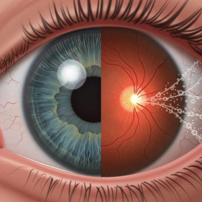
Pigment dispersion syndrome (PDS) is an eye condition in which microscopic particles of pigment from the iris become dislodged and accumulate within the eye’s drainage structures. This accumulation of pigment can obstruct the normal flow of fluid within the eye, potentially leading to elevated intraocular pressure and, in advanced cases, a condition known as pigmentary glaucoma. Understanding this condition is essential for individuals at risk, as early detection and management can help preserve vision and prevent serious complications.
What Happens When Pigment Breaks Free
The eye’s iris, the colored part responsible for controlling pupil size, contains specialized pigment cells that normally remain in place. In individuals with pigment dispersion syndrome, the anatomical structure of the eye causes the posterior surface of the iris to rub against the lens and its supporting structures, known as zonules. This friction gradually causes pigment granules to separate from the iris epithelium, the thin tissue layer covering the back of the iris.
Once released, these pigment particles circulate freely within the aqueous humor, the clear fluid that fills the front chamber of the eye. Rather than being naturally filtered out, many of these particles settle on various internal eye structures, including the corneal endothelium (the inner surface of the cornea), the trabecular meshwork (the eye’s drainage system), and the lens capsule. This deposition pattern creates distinctive clinical signs that ophthalmologists use to diagnose the condition.
The Role of Iris Shape in Pigment Release
The primary anatomical factor contributing to pigment dispersion syndrome is a concave curvature of the iris. Unlike a typical iris shape, individuals with PDS have an iris that bows backward toward the lens. This posterior bowing increases contact between the iris and the zonular fibers that support the lens, creating more friction during normal eye movements and pupil changes.
Several everyday activities can increase the rate of pigment shedding in susceptible individuals. During pupil dilation and constriction—which occurs when transitioning between bright and dark environments—the iris moves and flexes, generating friction against the lens zonules. Similarly, certain physical activities, particularly vigorous exercise, can accelerate pigment release by increasing the force of iris movement and aqueous fluid dynamics within the eye. Even routine activities like reading, blinking, and head movements can contribute to ongoing pigment liberation in eyes with the characteristic concave iris configuration.
How Pigment Accumulation Affects Eye Pressure
The trabecular meshwork functions as the eye’s primary drainage system, maintaining a delicate balance between fluid production and fluid outflow. When pigment granules accumulate excessively within this drainage network, they act as a physical obstruction, reducing the facility with which aqueous humor can exit the eye. Initially, this obstruction may cause only temporary increases in intraocular pressure (IOP), particularly following activities that trigger additional pigment shedding.
Over time, however, the chronic presence of pigment debris within the trabecular meshwork causes structural damage to the endothelial cells and supporting collagen beams that comprise this drainage system. These pathological changes result in permanently reduced drainage capacity, leading to sustained elevation of intraocular pressure. The longer the eye is exposed to elevated pressure, the greater the risk of damage to the optic nerve, the bundle of nerve fibers responsible for transmitting visual information to the brain.
Research indicates that individuals with pigment dispersion syndrome have approximately 15 times higher concentrations of pigment granules in their anterior chamber compared to people without the condition, reflecting the significant burden placed on the drainage system.
Distinctive Clinical Features and Signs
Ophthalmologists identify pigment dispersion syndrome through several characteristic findings observable during comprehensive eye examinations:
- Krukenberg spindle: A vertical band of pigment deposition on the inner surface of the cornea, created as pigment particles repeatedly strike the cornea during blinking and eye movements
- Trabecular meshwork pigmentation: Dense, homogeneous pigment accumulation visible within the drainage system when examined through specialized gonioscopic lenses
- Iris transillumination defects: Mid-peripheral areas of the iris that appear lighter than surrounding tissue when light is transmitted through the iris, indicating pigment loss from the epithelial layer
- Scheie stripe: Pigment deposition along the posterior lens capsule near the zonular attachment points, typically most visible in the lower portion of the eye
- Anterior iris pigment dusting: Fine pigment particles visible on the front surface of lighter-colored irises, resulting from debris settling on the iris surface
Progression to Pigmentary Glaucoma
Pigmentary glaucoma represents the advanced stage of pigment dispersion syndrome, occurring when elevated intraocular pressure causes measurable damage to the optic nerve. Not all individuals with pigment dispersion syndrome develop glaucoma; approximately 30 to 35 percent of patients with PDS experience sufficient pressure elevation to cause optic nerve damage. The progression from PDS to pigmentary glaucoma depends on multiple factors, including the severity of pigment accumulation, the drainage system’s inherent ability to handle excess pigment, and individual variations in optic nerve susceptibility to pressure-related injury.
Importantly, some patients develop pigmentary glaucoma despite never recording elevated eye pressure measurements during office visits. This phenomenon suggests that intermittent pressure spikes occurring outside clinical settings may be sufficient to cause progressive optic nerve damage in certain individuals.
Who Is Most Affected by This Condition
Pigment dispersion syndrome exhibits a clear demographic pattern in its prevalence. The condition is significantly more common in young adults, with onset typically occurring in the third and fourth decades of life. Caucasian populations experience higher rates of PDS compared to other ethnic groups. The condition demonstrates a pronounced male predominance, affecting men at considerably higher rates than women.
Myopia, or nearsightedness, is another important risk factor strongly associated with pigment dispersion syndrome. Individuals who are myopic have a substantially elevated risk of developing PDS compared to those with normal vision or hyperopia. This association suggests that the anatomical characteristics typical of myopic eyes—such as a longer axial length and different curvature patterns—may predispose to the posterior iris bowing that characterizes PDS.
The condition follows an autosomal dominant inheritance pattern, meaning that a single copy of the responsible genetic variant can cause the disease. Genetic studies have identified multiple different genetic loci associated with PDS, indicating that the condition results from mutations in various genes, each capable of producing the characteristic iris and drainage system abnormalities.
Symptoms Associated with Pressure Fluctuations
Many individuals with pigment dispersion syndrome experience no symptoms whatsoever, particularly in the early stages. The condition often goes undetected until identified incidentally during routine eye examinations or when symptoms of elevated pressure become apparent.
When symptoms do occur, they typically result from acute spikes in intraocular pressure rather than the baseline elevation of pressure. These episodic pressure increases can produce dramatic and distressing symptoms that resemble those of acute angle-closure glaucoma:
- Blurred or hazy vision
- Rainbow-colored halos surrounding light sources, caused by corneal edema from pressure elevation
- Eye discomfort or pain, ranging from mild aching to severe acute pain
- Headaches, often frontal or temporal in location
- Photophobia, or heightened sensitivity to bright light
- Redness of the eye
- Nausea, sometimes accompanied by vomiting in severe cases
These symptoms tend to be intermittent and may be triggered by specific activities. Physical exertion stands out as a particularly common trigger, as exercise simultaneously increases baseline intraocular pressure through increased aqueous humor production while also dislodging additional pigment through enhanced iris movement. Less strenuous activities, however, can also provoke symptoms in susceptible individuals. Transitions between light and dark environments, where pupil changes occur rapidly, frequently precipitate pressure spikes. Certain head postures and movements may also trigger episodes.
It is worth noting that pressure spikes produce more noticeable symptoms in eyes that already have a higher baseline intraocular pressure, as the additional pressure elevation from the spike represents a more substantial percentage change in overall eye pressure.
Diagnostic Evaluation and Testing
A comprehensive eye examination forms the foundation of pigment dispersion syndrome diagnosis. During the examination, the ophthalmologist performs several key assessments:
| Examination Component | What It Reveals |
|---|---|
| Slit lamp biomicroscopy | Krukenberg spindle, corneal findings, iris appearance, and anterior chamber pigmentation |
| Gonioscopy | Detailed visualization of trabecular meshwork pigmentation and drainage angle configuration |
| Tonometry | Intraocular pressure measurement to assess for elevation |
| Optic disc examination | Assessment for signs of optic nerve damage or cupping |
| Visual field testing | Detection of functional vision loss from glaucoma |
| Optical coherence tomography | High-resolution imaging of optic nerve structure and retinal nerve fiber layer thickness |
In some cases, provocative testing or dynamic monitoring may be warranted, particularly when symptoms suggest pressure spikes but routine measurements have not captured elevated readings. Some specialists may recommend baseline intraocular pressure measurement before and after exercise to document pressure response patterns in symptomatic individuals.
Management Strategies and Treatment Approaches
The management of pigment dispersion syndrome aims to prevent or slow the progression to pigmentary glaucoma by controlling intraocular pressure and minimizing ongoing pigment shedding. Treatment approaches vary based on whether the patient has developed glaucomatous optic nerve damage and the severity of pressure elevation.
For individuals with pigment dispersion syndrome but normal intraocular pressures and no optic nerve damage, conservative observation is typically recommended. Regular monitoring with serial intraocular pressure measurements, optic disc assessment, and visual field testing at intervals appropriate to the individual’s risk profile allows early detection of glaucoma development before significant vision loss occurs.
When intraocular pressure becomes elevated or shows a pattern of significant fluctuations, medication to lower pressure may be prescribed. Topical medications that reduce aqueous humor production or increase aqueous outflow are typically initiated as first-line therapy. For individuals who develop episodes of acute pressure elevation associated with symptoms, prophylactic medication before known trigger activities—such as strenuous exercise—may be recommended.
Laser interventions can provide an alternative or adjunctive approach to pressure management. Peripheral iridotomy, a procedure that creates a small opening in the iris, may reduce pressure spikes in some patients by altering aqueous flow dynamics. Laser treatment directed at the trabecular meshwork to increase drainage facility represents another option when medications are insufficient or poorly tolerated.
For individuals with advanced pigmentary glaucoma unresponsive to medical and laser therapy, incisional surgical procedures that create alternative aqueous drainage pathways may be necessary to prevent further optic nerve damage and vision loss.
Lifestyle modifications also form an important component of management. Individuals at high risk or with established disease may benefit from avoiding strenuous exercise or modifying exercise intensity, though the evidence for exercise-related pressure spikes is variable across the population. Maintaining consistent use of prescribed medications, protecting eyes from bright light exposure when problematic, and maintaining good overall eye health are universally recommended.
Long-Term Vision Prognosis
The long-term outlook for individuals with pigment dispersion syndrome depends significantly on whether the condition progresses to pigmentary glaucoma and how aggressively the elevated intraocular pressure is managed. Early detection and appropriate treatment substantially reduce the risk of significant vision loss. However, advanced pigmentary glaucoma that goes undetected or untreated can result in progressive peripheral vision loss and, in severe cases, blindness.
As patients age, the rate of pigment dispersion typically decreases, likely due to exhaustion of the available pigment stores and potential changes in iris-zonule contact geometry over time. However, accumulated damage to the trabecular meshwork and established glaucomatous optic nerve damage persist, requiring continued pressure management to prevent further deterioration.
Frequently Asked Questions
Can pigment dispersion syndrome be prevented?
Since pigment dispersion syndrome is determined by inherent eye anatomy and genetics, it cannot be prevented. However, in individuals with diagnosed PDS, minimizing pigment shedding through avoiding strenuous activity and managing intraocular pressure can help prevent progression to glaucoma.
Is pigment dispersion syndrome the same as pigmentary glaucoma?
No, they represent different points along the same disease spectrum. Pigment dispersion syndrome is characterized by pigment liberation and accumulation without necessarily causing optic nerve damage or elevated pressure. Pigmentary glaucoma occurs when PDS results in pressure elevation significant enough to damage the optic nerve.
How often should individuals with PDS have eye examinations?
The frequency of monitoring depends on individual risk factors and whether glaucoma has developed. Some specialists recommend annual evaluations for patients with confirmed PDS, with more frequent visits for those showing pressure elevation or signs of optic nerve damage.
Can vision loss from pigmentary glaucoma be reversed?
Unfortunately, vision loss from glaucoma cannot be reversed. This underscores the importance of early detection and prevention of progression through adequate pressure control.
References
- Pigmentary Glaucoma and Pigment Dispersion Syndrome — American Academy of Ophthalmology. 2024. https://glaucoma.org/types/pigmentary-glaucoma
- Pigment Dispersion Syndrome — Columbia University Vagelos College of Physicians and Surgeons, Department of Ophthalmology. 2024. https://www.vagelos.columbia.edu/departments-centers/ophthalmology
- Pigmentary Glaucoma and Pigment Dispersion Syndrome — EyeWiki, American Academy of Ophthalmology. 2024. https://eyewiki.org/Pigmentary_Glaucoma_and_Pigment_Dispersion_Syndrome
- Pigment Dispersion Syndrome: Symptoms, Causes, and Treatment — Medical News Today. 2024. https://www.medicalnewstoday.com/articles/what-is-pigment-dispersion-syndrome
- Pigment Dispersion Syndrome and Pigmentary Glaucoma — All About Vision. 2024. https://www.allaboutvision.com/conditions/pigment-dispersion-syndrome/
- Pigment Dispersion Syndrome and Pigmentary Glaucoma: The Basics — EyeWorld. 2025. https://www.eyeworld.org/2025/pigment-dispersion-syndrome-and-pigmentary-glaucoma-the-basics/
- Pigment Dispersion Syndrome — Genetic and Rare Diseases Information Center (GARD), National Institutes of Health. 2024. https://rarediseases.info.nih.gov/diseases/4356/pigment-dispersion-syndrome
Similar Articles


Read full bio of Sneha Tete