Pilar Sheath Acanthoma Pathology
Detailed histopathological analysis of pilar sheath acanthoma, a benign follicular tumour with distinctive dermal lobular proliferation.

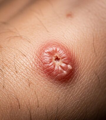
Pilar sheath acanthoma is a rare benign neoplasm derived from the outer root sheath of the hair follicle, most commonly presenting as a small papule on the skin of the upper lip with a central pore. This condition is characterized by a lobular proliferation of squamous epithelium extending into the dermis, distinguishing it from other follicular tumours.
Introduction
Pilar sheath acanthoma, first described by Mehregan and Brownstein in 1978, represents a benign follicular hamartoma that typically affects middle-aged to elderly adults. It manifests clinically as a solitary, skin-coloured papule or nodule, 3–10 mm in diameter, often featuring a central keratin-filled pore or depression from which keratin plugs can be expressed. While the upper lip is the classic site, lesions may occasionally appear on other facial areas such as the cheeks, lower lip, or rarely the eyebrow. Histopathologically, it is defined by a central dilated follicular infundibulum connected to the surface, surrounded by radiating lobules of epithelial cells showing outer root sheath differentiation. This article delves into the microscopic features, aiding dermatopathologists in accurate diagnosis and differentiation from mimics like dilated pore of Winer or trichofolliculoma.
Clinical features
Clinically, pilar sheath acanthoma presents as a small, asymptomatic, skin-coloured papule with a central pore, predominantly on the upper lip. The lesion measures up to 5–10 mm in diameter and may express keratin debris upon squeezing, but no hair emerges from the pore. It is usually solitary and does not associate with systemic disorders, though rare multiple lesions have been reported. In atypical cases, such as on the eyebrow, the central opening may not be clinically apparent. Differential clinical diagnoses include basal cell carcinoma, epidermoid cyst, trichoepithelioma, and dilated pore of Winer. Biopsy is essential for confirmation, as the benign nature is only verified histologically.
- Solitary papule or nodule, 3–10 mm diameter
- Skin-coloured with central 1–2 mm pore or depression
- Predilection for upper lip in adults over 40 years
- Asymptomatic; keratin expressible but no hair growth
- Rare sites: cheeks, chin, eyebrow
Histology
At low magnification, pilar sheath acanthoma displays a lobular proliferation of benign squamous epithelium within the dermis, centred around small cystic spaces that connect to the epidermal surface via a dilated pore filled with keratinous debris (figures 1 and 2). These epithelial lobules radiate outward from the central cystic cavity, extending into the deep dermis or even subcutis, composed of bland keratinocytes without atypia or mitoses.
The lining of the cystic spaces varies: some exhibit a prominent granular layer akin to epidermoid cysts (figure 3), while others show an attenuated granular layer resembling trichilemmal cysts (figure 4). Clear cells with glycogen-rich cytoplasm are frequently observed within the lobules (figures 5 and 6), indicative of outer root sheath differentiation. An eosinophilic basement membrane often surrounds the lobules, further supporting follicular sheath origin. No fibrovascular stroma or hair shaft formation is present, distinguishing it from other adnexal tumours.
| Feature | Description |
|---|---|
| Architecture | Lobular epithelial proliferation around central cystic infundibulum opening to surface |
| Cystic spaces | Small, keratin-filled; variable granular layer (epidermoid or trichilemmal type) |
| Cells | Bland squamous keratinocytes; clear cells with glycogen |
| Stroma | Minimal; eosinophilic basement membrane around lobules |
| Depth | Dermis to subcutis; no atypia or mitoses |
Differential diagnosis
Pilar sheath acanthoma must be differentiated from several benign follicular lesions based on architectural patterns and cytological features.
Dilated pore of Winer
Dilated pore of Winer features a larger central cystic space lined by acanthotic epithelium with thin, radiating epithelial strands or finger-like projections into the surrounding stroma, contrasting the thicker, more compact lobules of pilar sheath acanthoma. The cystic dilatation is more prominent, and lobular proliferation is absent.
Trichofolliculoma
Trichofolliculoma shows immature hair follicles radiating from a central cystic structure, often with well-formed fibrovascular stroma and occasional hair shafts. In pilar sheath acanthoma, abortive follicle-like structures lack maturation, and stroma is inconspicuous.
Trichilemmoma
Trichilemmoma exhibits peripheral palisading and more uniform clear cell change without the central cystic connection or lobular arrangement around keratin-filled spaces.
Epidermoid cyst
Epidermoid cysts lack the radiating lobules and show a single large cystic cavity with uniform epidermoid keratinization, without dermal extension.
- Dilated pore: Thin radiating strands, large cyst
- Trichofolliculoma: Mature follicles, stroma present
- Trichilemmoma: Palisading, no central pore
- Other: Rule out BCC (atypia) or basosquamous lesions
Management
As a benign neoplasm, pilar sheath acanthoma requires no treatment unless for cosmetic concerns or functional impairment. Excisional biopsy or shave excision is curative and diagnostic. Small lesions can be removed via punch biopsy (2 mm) with suture closure. Curettage and electrodesiccation are alternatives for superficial lesions. Recurrence is rare post-excision.
Frequently asked questions
What is pilar sheath acanthoma?
A benign hair follicle sheath tumour presenting as a small facial papule with central pore.
Where does it commonly occur?
Primarily on the upper lip of middle-aged/elderly adults.
How is it diagnosed?
By histopathology showing dermal lobules of squamous epithelium around a central keratin-filled cyst.
Is treatment necessary?
No, unless cosmetic; excision is curative.
What does it look like under the microscope?
Lobular proliferation with clear cells, eosinophilic membrane, variable granular layer.
Prognosis
Pilar sheath acanthoma is entirely benign with no malignant potential or syndromic associations. Post-excision, no recurrence or follow-up is needed.
References
- Pilar sheath acanthoma — Wikipedia. 2023. https://en.wikipedia.org/wiki/Pilar_sheath_acanthoma
- Pilar sheath acanthoma pathology — DermNet NZ. 2023. https://dermnetnz.org/topics/pilar-sheath-acanthoma-pathology
- An Unusual Location of a Pilar Sheath Acanthoma — PMC (Indian Dermatol Online J). 2016-01-29. https://pmc.ncbi.nlm.nih.gov/articles/PMC4738487/
- Pilar Sheath Acanthoma — MalaCards. 2023. https://www.malacards.org/card/pilar_sheath_acanthoma
- Pilar Sheath Acanthoma — Perri Dermatology. 2011-06-26. https://perridermatology.com/dr-perris-blog/hair-follicle-neoplasms-pilar-sheath-acanthoma/
- Pilar sheath acanthoma — VisualDx. 2023. https://www.visualdx.com/visualdx/diagnosis/pilar+sheath+acanthoma?diagnosisId=54715&moduleId=101
- Pilar sheath acanthoma — DermNet NZ. 2013. https://dermnetnz.org/topics/pilar-sheath-acanthoma
Similar Articles


Read full bio of medha deb


