Pityriasis Rotunda Explained: Symptoms, Diagnosis & Treatment
Rare skin disorder with circular scaly patches linked to malignancy and systemic diseases.

Author: Dermatologist Review Team Synonym(s): Pityriasis circinata et marginata, Rotund pityriasis
What is pityriasis rotunda?
Pityriasis rotunda is a rare chronic disorder of keratinization characterised by the development of well-defined round or oval scaly patches that may be hypo or hyperpigmented. It is a type of ichthyosiform dermatosis resembling tinea corporis but without inflammatory changes or infectious aetiology. Lesions are typically asymptomatic and persistent, distinguishing them from resolving conditions like pityriasis rosea.
The condition was first described in 1908 by Bertram in Japanese patients and later recognised globally. It manifests in two types: Type I (paraneoplastic, multiple lesions in adults) strongly associated with internal malignancy, and Type II (idiopathic, fewer lesions, often in younger patients or those with non-malignant systemic diseases).
Who gets pityriasis rotunda?
Pityriasis rotunda exhibits distinct epidemiological patterns. Type I predominantly affects adults over 40-60 years, with higher prevalence in individuals of Asian or African descent. It is rare in Caucasians. Multiple lesions (>10) signal high risk of underlying malignancy in 60-100% of cases.
Type II occurs in younger patients, including children, and is linked to non-neoplastic conditions. Familial cases are exceptional. Overall incidence is low, estimated at fewer than 100 reported cases worldwide, though underdiagnosis may occur due to asymptomatic nature.
- High-risk groups: Elderly Asians/Africans (Type I)
- Systemic associations: Common in Japan, South Africa
- Gender: Slight female predominance in some series
Clinical features of pityriasis rotunda
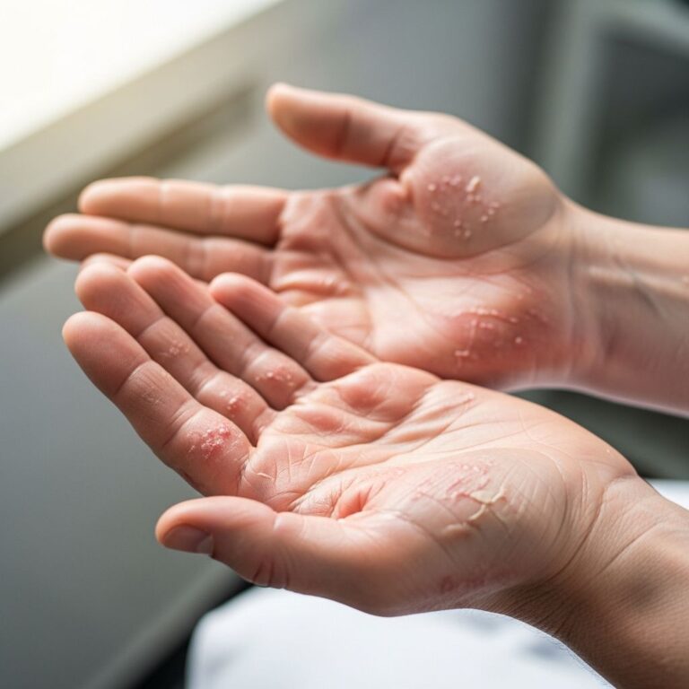
Lesions present as 2-10 cm diameter, perfectly circular or oval, sharply demarcated plaques with fine ichthyosiform scale and adherent collarette at the edge. Central clearing may occur in larger patches. They lack erythema, induration, or symptoms like itch/pruritus, persisting for years without spontaneous resolution in most cases.
Distribution favours trunk (back, buttocks, abdomen), proximal extremities; spares flexures, face, scalp, mucous membranes. Multiple coalescing lesions form polycyclic patterns over buttocks/thighs. Hyperpigmented in darker skin types; hypopigmented variants rare.
| Feature | Type I (Paraneoplastic) | Type II (Idiopathic) |
|---|---|---|
| Age of onset | >40-60 years | Any age, often younger |
| Number of lesions | >10, widespread | <10, localised |
| Malignancy risk | High (hepatocellular carcinoma, gastric ca) | Low/none |
| Associations | Chronic illness | Systemic non-malignant disease |
Pathogenesis of pityriasis rotunda
Aetiology remains unclear. No infectious cause despite tinea-like morphology; KOH scrapings consistently negative. Proposed links to keratinocyte differentiation defects: reduced expression of filaggrin, loricrin, profilaggrin-N terminus, and filaggrin 2 observed in lesional epidermis, akin to ichthyosis vulgaris.
Type I likely paraneoplastic; cytokines/neuropeptides from malignancy may induce epidermal changes. Type II may represent acquired ichthyosis variant. Genetic factors unproven; sporadic cases predominate.
Differential diagnosis of pityriasis rotunda
Key differentials include infectious and inflammatory mimics:
- Tinea corporis: Annular with central clearing, positive KOH, responds to antifungals
- Pityriasis versicolor: Hypopigmented, fine scale, Malassezia on microscopy
- Granuloma annulare: Annular plaques with central atrophy, palpable
- Erythema chronicum migrans: Expansile post-tick bite, history/symptoms
- Acquired ichthyosis: Diffuse scaling, malignancy association
- Nummular eczema: Vesicles, itch, oozing
Biopsy differentiates; clinical context (age, number lesions, systemic symptoms) guides suspicion.
Investigations for pityriasis rotunda
Mandatory screening for internal disease, especially Type I:
- Full history/exam: Weight loss, systemic symptoms
- Laboratory: FBC, LFTs, TFTs, glucose, inflammatory markers, tumour markers (AFP for HCC)
- Imaging: Chest X-ray, abdominal US/CT, endoscopy if indicated
- Skin scrapings: KOH for fungus
- Biopsy: Confirm diagnosis, rule out mycosis fungoides
Multiple lesions in older adults warrant urgent malignancy workup.
Histology of pityriasis rotunda
Characteristic but non-specific: basket-weave orthokeratosis ± focal parakeratosis, absent/reduced granular layer, elongated rete ridges, mild basal hyperpigmentation, pigment incontinence, superficial perivascular lymphocytic infiltrate. Follicular plugging common. No atypia or malignancy.
Treatment of pityriasis rotunda
No specific cure. Lesions often resolve with treatment of underlying malignancy/systemic disease. Symptomatic topicals largely ineffective:
- Emollients, keratolytics (salicylic/lactic acid): Minimal benefit
- Topical steroids, antifungals, tars: No response
- Topical retinoids (tazarotene), oral retinoids: Limited success
- Calcipotriol (vitamin D3 analogue): Gradual improvement in select cases
Observation appropriate for asymptomatic lesions; monitor for progression/resolution. Multidisciplinary approach essential.
Complications of pityriasis rotunda
- Malignancy: Type I marker (HCC 30-50%, gastric ca)
- Cosmesis from persistent hypopigmentation/hyperpigmentation
- Diagnostic delay if misdiagnosed as infection
Prevention of pityriasis rotunda
Non-preventable. Early malignancy screening in at-risk populations (elderly with multiple lesions) may indirectly benefit.
Frequently asked questions about pityriasis rotunda
Is pityriasis rotunda contagious?
No. Despite tinea-like appearance, scrapings are negative and it is not infectious.
Does pityriasis rotunda always mean cancer?
No, but multiple lesions in older adults have high (>50%) malignancy association requiring urgent investigation.
Can pityriasis rotunda be treated with antifungals?
No. Responds poorly; treat underlying cause if identified.
Will pityriasis rotunda go away on its own?
Rarely; persistent for years but may resolve post-malignancy treatment.
What does pityriasis rotunda look like?
Round/oval 2-10cm scaly plaques, trunk/extremities, no redness/itch.
References
- Pityriasis rotunda — DermNet NZ. 2023. https://dermnetnz.org/topics/pityriasis-rotunda
- Pityriasis Rotunda | Treatment & Management — StatPearls [Internet]. 2024-05-01. https://www.statpearls.com/point-of-care/27256
- Pityriasis rotunda and hyperprolactinemia — Actas Dermo-Sifiliográficas. 2016-07-01. http://www.actasdermo.org/en-pityriasis-rotunda-hyperprolactinemia-articulo-S1578219016300828
- Pityriasis rotunda — Indian Journal of Dermatology, Venereology and Leprology. 2005. https://ijdvl.com/pityriasis-rotunda/
- Pityriasis Rotunda — MD Searchlight. 2024. https://mdsearchlight.com/skin-problems-and-treatments/pityriasis-rotunda/
- Pityriasis rotunda — VisualDx. 2024. https://www.visualdx.com/visualdx/diagnosis/pityriasis+rotunda?diagnosisId=55824&moduleId=101
Similar Articles


Read full bio of medha deb