Porokeratosis Of Mibelli: Causes, Diagnosis, Treatment Guide
Comprehensive guide to Porokeratosis of Mibelli: symptoms, causes, diagnosis, treatment, and risk of malignancy.

Porokeratosis of Mibelli is a rare dermatological disorder characterized by the development of annular plaques with a distinctive hyperkeratotic ridge-like border, known as the cornoid lamella on histopathology. This condition represents the second most common form of porokeratosis and typically manifests in childhood or young adulthood.
Introduction
Porokeratosis encompasses a group of disorders of keratinization presenting as annular lesions with a raised, thread-like border. Porokeratosis of Mibelli, named after Vittorio Mibelli who first described it in 1893, features lesions that begin as small brown papules evolving into plaques with irregular outlines and a characteristic peripheral furrow. The condition arises from clonal expansion of abnormal keratinocytes, leading to faulty keratinization.
Demographics
Porokeratosis of Mibelli predominantly affects children, adolescents, and young adults, with onset sometimes linked to immunosuppression. It exhibits a male predominance, occurring twice as frequently in males as in females. While often sporadic, familial cases with autosomal dominant inheritance have been documented. The prevalence is low, classified as a rare disease by Orphanet.
Causes
The precise etiology remains unclear, but lesions originate from localized proliferation of a keratinocyte clone with keratinization defects. Key contributing factors include:
- Genetic mutations: Mutations in the mevalonate kinase gene (MVK) have been implicated.
- Immunosuppression: Common in organ transplant recipients on immunosuppressive therapy, with onset 3 weeks to 14 years post-initiation.
- UV radiation: Sun exposure exacerbates and may induce lesions.
- Drugs and therapies: Associations with phototherapy, radiotherapy, and biologics.
These triggers disrupt normal epidermal turnover, culminating in parakeratotic column formation.
Clinical features
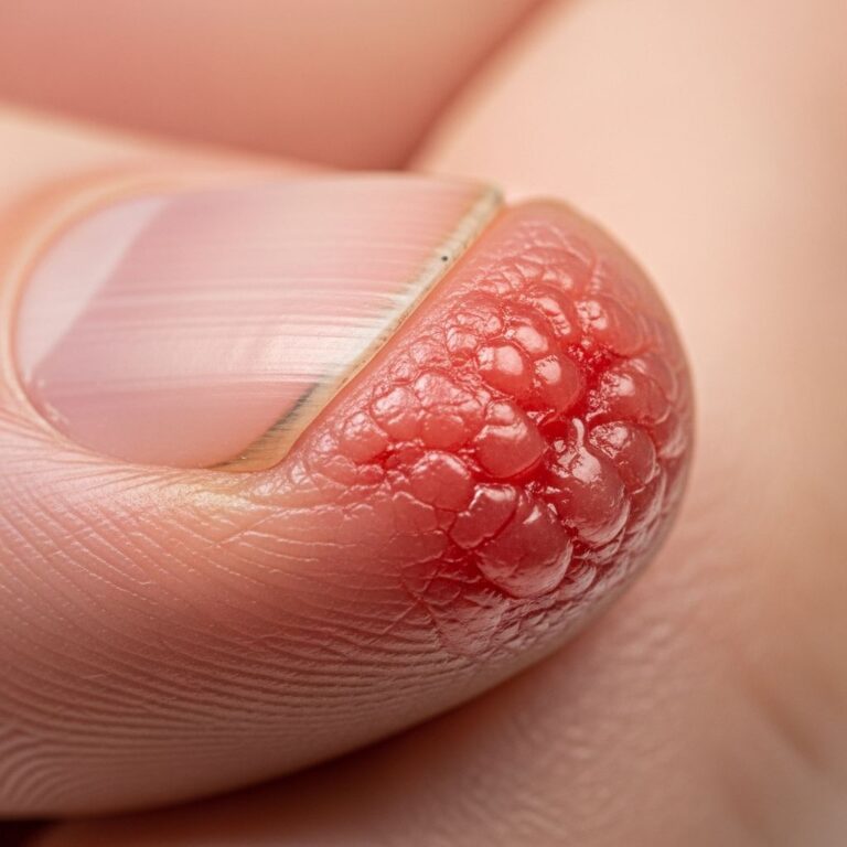
Lesions initiate as small, light brown, scaly papules that coalesce into plaques with irregular borders and a warty, hyperkeratotic rim featuring a central furrow. Plaques vary from a few centimeters to giant forms exceeding 20 cm. Common sites include extremities (hands, feet, lower legs), but trunk, neck, face, genitals, palms, soles, and rarely mucosae may be involved. Lesions are typically asymptomatic but may itch occasionally; they wax and wane over years.
| Feature | Description |
|---|---|
| Appearance | Brownish, annular plaques with keratotic thread-like border |
| Size | 1-20 cm; usually small but can be giant |
| Locations | Limbs (esp. distal), trunk, genitals; rarely face/mucosa |
| Symptoms | Asymptomatic or pruritic; chronic progression |
Complications
The primary concern is malignant transformation in approximately 7-10% of cases, typically to squamous cell carcinoma (most common), or basal cell carcinoma. Risk escalates with longstanding lesions, UV exposure, and immunosuppression. Rare fatalities from metastatic SCC reported. Chronic lesions impair quality of life cosmetically and psychologically.
Diagnosis
Diagnosis relies on clinical morphology and confirmed by skin biopsy revealing the pathognomonic cornoid lamella: a narrow vertical stack of parakeratotic cells over an epidermal depression. Dermoscopy shows the white thread-like rim with furrows.
Differential diagnoses include:
- Actinic keratosis
- Elastosis perforans serpiginosa
- Annular lichen planus
- Psoriasis
- Bowen’s disease
- Circumscribed palmoplantar hypokeratosis
- Focal palmoplantar keratoderma
Differential diagnoses
Key distinctions:
| Condition | Distinguishing Features |
|---|---|
| Actinic keratosis | Sun-damaged skin; no annular form or cornoid lamella |
| Psoriasis | Well-defined plaques without peripheral ridge; silvery scales |
| Bowen’s disease | Scaly red plaque; lacks keratotic rim; biopsy differentiates |
| Lichen planus | Polygonal papules; violaceous hue; Wickham striae on dermoscopy |
Treatment
No curative therapy exists; treatments aim to alleviate symptoms, improve cosmesis, and prevent progression. Recurrence is common.
Conservative measures:
- Sun protection (critical to reduce malignancy risk)
- Regular dermatologic surveillance for malignant change
Topical therapies:
- 5-Fluorouracil (5-FU) cream
- Imiquimod cream (effective for classical Mibelli)
- Retinoids, vitamin D analogs, diclofenac gel
Procedural interventions:
- Cryotherapy with liquid nitrogen
- Laser ablation (CO2, fractional)
- Photodynamic therapy (PDT)
- Dermabrasion, curettage
- Surgical excision (for small lesions or suspected malignancy)
Suspected skin cancers require biopsy or excision.
Outcome
Porokeratosis of Mibelli follows a chronically progressive course with periods of stability and expansion. Lesions persist indefinitely, impacting quality of life aesthetically. Malignancy risk necessitates lifelong monitoring, especially in high-risk patients (immunosuppressed, sun-exposed). Early intervention and sun avoidance mitigate complications.
Frequently Asked Questions
Q: Is Porokeratosis of Mibelli contagious?
A: No, it is not infectious; it results from intrinsic keratinocyte abnormalities, not pathogens.
Q: Can Porokeratosis of Mibelli turn into cancer?
A: Yes, 7-10% of lesions may develop into squamous cell carcinoma or basal cell carcinoma, particularly longstanding ones.
Q: What is the best treatment for Porokeratosis of Mibelli?
A: No single best treatment; options like imiquimod, cryotherapy, or excision are used based on lesion size/location. Recurrence is frequent.
Q: Does sun exposure worsen Porokeratosis of Mibelli?
A: Yes, UV radiation promotes lesion progression and malignancy risk; rigorous sun protection is essential.
Q: Is Porokeratosis of Mibelli hereditary?
A: Often sporadic, but autosomal dominant inheritance occurs in some families, linked to MVK mutations.
References
- Porokeratosis of Mibelli — Orphanet. 2020-08-01. https://www.orpha.net/en/disease/detail/735
- Understanding Porokeratosis: Causes, Symptoms & Effective Treatments — MDCS Dermatology. 2023-01-15. https://www.mdcsnyc.com/post/understanding-porokeratosis-causes-symptoms-effective-treatments
- Porokeratosis of Mibelli — DermNet NZ. 2023-05-20. https://dermnetnz.org/topics/porokeratosis-of-mibelli
- Porokeratosis — MD Searchlight. 2024-02-10. https://mdsearchlight.com/skin-problems-and-treatments/porokeratosis/
- What Is Porokeratosis — WebMD. 2024-07-12. https://www.webmd.com/skin-problems-and-treatments/what-is-porokeratosis
- Porokeratosis: Types, causes, treatments, and pictures — Medical News Today. 2023-11-05. https://www.medicalnewstoday.com/articles/porokeratosis
- Porokeratosis — StatPearls, NCBI Bookshelf. 2023-04-22. https://www.ncbi.nlm.nih.gov/books/NBK532290/
Similar Articles


Read full bio of Sneha Tete