Porokeratosis Pathology: Guide To Cornoid Lamella And Subtypes
Detailed histopathological examination and clinical insights into porokeratosis, a keratinization disorder with cornoid lamella.

Porokeratosis represents a spectrum of rare skin disorders characterized by abnormal keratinization, distinguished histologically by the hallmark
cornoid lamella
—a column of parakeratotic cells in the stratum corneum overlying an epidermal invagination. This condition arises from clonal keratinocyte proliferation, often linked to genetic mutations in the mevalonate-isoprenoid pathway, leading to diverse clinical presentations from localized plaques to disseminated lesions. Understanding its pathology is crucial for accurate diagnosis and monitoring for potential malignant transformation.Introduction
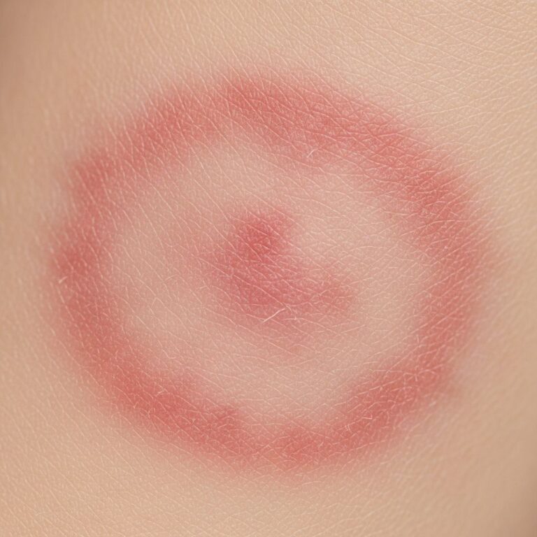
Porokeratosis encompasses heterogeneous acquired or congenital keratinizing disorders of unknown etiology, primarily featuring faulty epidermal keratinization due to abnormal clonal expansion of keratinocytes. First described in 1893, it includes over nine subtypes, with lesions presenting as well-demarcated annular papules or plaques with an atrophic center and hyperkeratotic ridge. The unifying microscopic feature, the cornoid lamella, correlates clinically with the raised, scaly border observed in all variants. Genetic predispositions, UV exposure, and immunosuppression contribute to its pathogenesis, with a noted risk of squamous cell carcinoma (SCC) development.
Histopathology
The defining histopathological hallmark of porokeratosis is the
cornoid lamella
, an invaginated column of parakeratotic cells (retaining nuclei in the stratum corneum) within the epidermis, surrounded by hypogranulosis or agranulosis and dyskeratosis. This structure represents proliferating keratinocytes that fail to mature fully, resulting in unilateral thinning of the stratum corneum centrally and hyperkeratosis peripherally. Additional findings include:- Absence of the granular layer beneath the lamella.
- Focal epidermal necrosis or blistering (rare).
- Pigment incontinence and dermal melanophages in pigmented forms.
- Interface dermatitis or perivascular mononuclear infiltrates in some variants like prurigo nodularis-like DSAP.
In dermoscopy, the cornoid lamella appears as a hyperkeratotic white, yellow, or brown peripheral edge (double-track sign), with a homogeneous, scar-like central area possibly containing brown globules or vascular structures. Ink-enhanced dermoscopy aids differentiation by highlighting the ridge-like lamella after alcohol cleaning. Reflectance confocal microscopy reveals similar features, facilitating non-invasive diagnosis.
Clinical Subtypes and Their Pathology
Porokeratosis manifests in multiple subtypes, each with distinct distributions, triggers, and histopathological nuances, though all share the cornoid lamella.
Classic Porokeratosis of Mibelli
This form typically affects extremities in infants or children, presenting as annular plaques with a raised keratotic border and atrophic, hypopigmented center. Histology shows prominent cornoid lamella with central hairless atrophy; malignancy risk is elevated in longstanding lesions.
Disseminated Superficial Actinic Porokeratosis (DSAP)
The most common variant, DSAP features numerous small, annular lesions on sun-exposed areas like legs, exacerbated by UV light. Autosomal dominant with reduced penetrance, it shows fewer lesions in treated areas with diclofenac gel (up to 53.8% reduction). Pathology includes thin cornoid lamella over sun-damaged skin, with postinflammatory hyperpigmentation in skin of color.
Linear Porokeratosis
Following Blaschko’s lines, this congenital form develops in infancy, with zosteriform or systematized patterns. Histology mirrors classic types but along linear distributions; high SCC risk reported.
Disseminated Superficial Porokeratosis (DSP)
Affects non-sun-exposed skin, sparing the face, with widespread lesions. Often linked to immunosuppression.
Other Variants
- Porokeratosis Palmaris et Plantaris Disseminata: Confined to palms/soles.
- Eruptive Disseminated Porokeratosis (EDP): Extremely pruritic, self-resolving with hyperpigmented patches.
- Giant Porokeratosis: Large plaques >5 cm, higher malignancy potential.
| Subtype | Distribution | Triggers | Key Pathology |
|---|---|---|---|
| Mibelli | Extremities | Genetic | Prominent lamella, atrophy |
| DSAP | Sun-exposed | UV | Thin lamella, pigmentation |
| Linear | Blaschko lines | Congenital | Linear lamella |
| DSP | Trunk/limbs | Immunosuppression | Widespread lamella |
Pathogenesis and Genetics
Porokeratosis stems from defects in the
mevalonate-isoprenoid pathway
, essential for cholesterol synthesis and keratinocyte differentiation. Mutations in genes like MVK, PMVK, MVD, FDPS, and others cause abnormal prenylation of proteins, leading to clonal keratinocyte expansion. Autosomal dominant inheritance predominates in DSAP and Mibelli types, with somatic mosaicism in linear forms. UV radiation and immunosuppression exacerbate lesions by promoting proliferation.Diagnosis
Diagnosis relies on clinical morphology, dermoscopy (double-track sign, radial vessels), and biopsy confirming cornoid lamella. Differentials include actinic keratosis, SCC, nummular dermatitis, and tinea corporis. In skin of color, lesions appear violaceous-brown with accentuated scaly ridges. Advanced imaging like reflectance confocal microscopy shows parakeratosis and spongiosis non-invasively.
Complications
The primary concern is
malignant transformation
to SCC (most common) or basal cell carcinoma, especially in larger, longstanding lesions on sun-exposed sites. Risk factors include immunosuppression and giant forms; regular monitoring is essential.Treatment
No cure exists; treatments aim at symptom relief and lesion reduction. Options include:
- Topical: Diclofenac 3% gel (53.8% lesion reduction at 3 months), imiquimod, retinoids.
- Photodynamic Therapy (PDT): MAL-PDT improves texture/color, superior to cryotherapy in some series.
- Physical: Cryotherapy, curettage, laser (CO2, Er:YAG).
- Systemic: Acitretin for disseminated forms.
EDP often resolves spontaneously. Targeting the isoprenoid pathway with statins shows promise in research.
Frequently Asked Questions (FAQs)
What is the hallmark feature of porokeratosis on histopathology?
The cornoid lamella—a parakeratotic column over epidermal invagination.
Is porokeratosis hereditary?
Yes, many subtypes like DSAP are autosomal dominant.
Can porokeratosis turn into cancer?
Yes, particularly SCC in longstanding lesions; monitor regularly.
How is porokeratosis diagnosed?
Via clinical exam, dermoscopy, and biopsy.
What treatments work best for DSAP?
Diclofenac gel and PDT show good efficacy.
Conclusion
Porokeratosis pathology hinges on the cornoid lamella, reflecting underlying genetic and pathway defects. Early recognition via dermoscopy and biopsy, coupled with vigilant monitoring for malignancy, guides management in this enigmatic group of disorders.
References
- Porokeratosis: A Review of its Pathophysiology, Clinical… — Actas Dermo-Sifiliográficas. 2020-10-15. https://www.actasdermo.org/en-porokeratosis-a-review-its-pathophysiology-articulo-S1578219020302055
- Porokeratosis — HCA Healthcare Scholarly Commons. 2023. https://scholarlycommons.hcahealthcare.com/cgi/viewcontent.cgi?article=1682&context=hcahealthcarejournal
- Porokeratoses—A Comprehensive Review on the Genetics and… — PMC/NCBI. 2023-12-01. https://pmc.ncbi.nlm.nih.gov/articles/PMC10744643/
- Porokeratosis: overview, clinical subtypes, histology… — YouTube (Strong Medicine). 2020. https://www.youtube.com/watch?v=pEodUB6UD0Y
- Porokeratosis — DermNet NZ. 2023. https://dermnetnz.org/topics/porokeratosis
Similar Articles


Read full bio of medha deb