Posterior Pituitary: What It Is & Function
Understanding the posterior pituitary: anatomy, hormones, and vital functions.

Understanding the Posterior Pituitary: Structure and Function
The posterior pituitary, also known as the neurohypophysis, is a small but essential component of your endocrine system. Located at the base of your brain, this gland plays a crucial role in regulating several vital bodily functions. Unlike many other glands in the body, the posterior pituitary does not produce hormones itself. Instead, it stores and releases two critical hormones produced by the hypothalamus: oxytocin and vasopressin (antidiuretic hormone or ADH). Understanding how this gland works provides insight into how your body maintains homeostasis and manages essential processes like water balance, lactation, and uterine contraction.
Anatomy and Location of the Posterior Pituitary
Your pituitary gland is a remarkable structure despite its tiny size. Measuring only about one-third of an inch in diameter—roughly the size of a pea—this gland sits in a special bony cavity called the sella turcica, located at the base of your brain directly beneath the hypothalamus and behind the bridge of your nose. The pituitary gland consists of two distinct lobes that work together: the posterior pituitary (the back lobe) and the anterior pituitary (the front lobe).
The posterior pituitary is the smaller of the two lobes, making up approximately 20% of the total weight of the pituitary gland, while the anterior pituitary comprises about 80%. Despite its smaller size, the posterior pituitary’s functions are absolutely essential for human health and survival. The gland is connected to the hypothalamus through a specialized stalk-like structure called the infundibular stalk, which contains blood vessels and nerve cells. This connection is vital because it allows direct communication between the hypothalamus and the posterior pituitary through both neural signals and hormonal pathways.
Cellular Structure and Composition
The posterior pituitary has a unique cellular composition that distinguishes it from the anterior pituitary. Functionally, the posterior pituitary acts as an extension of the hypothalamus and consists of specialized neurosecretory tissue derived from the embryonic forebrain. The primary cell types found in the posterior pituitary include:
Magnocellular Neurosecretory Cells: These neurons originate in specific regions of the hypothalamus—the supraoptic nuclei and paraventricular nuclei—and extend their axons (nerve projections) down through the infundibular stalk to the posterior pituitary. These specialized neurons are responsible for synthesizing and transporting the hormones oxytocin and vasopressin.
Pituicytes: These are specialized glial cells that resemble astrocytes found in the brain. Pituicytes play a supportive role in assisting with the storage and release of neurohypophysial hormones.
Herring Bodies: Within the posterior pituitary, hormones are stored in specialized neurosecretory structures called Herring bodies. These are located at nerve terminals and serve as storage depots for oxytocin and antidiuretic hormone until they are needed for release into the bloodstream.
The Two Hormones of the Posterior Pituitary
The posterior pituitary stores and releases exactly two hormones, both synthesized in the hypothalamus. These hormones have distinct roles and functions throughout the body:
Antidiuretic Hormone (ADH) or Vasopressin
Antidiuretic hormone, commonly referred to as vasopressin, is synthesized in the supraoptic nuclei of the hypothalamus. This hormone functions as a water-preserving agent in the body. ADH regulates how much water your kidneys retain, which directly affects your hydration status and blood pressure. When your body experiences dehydration or an increase in blood osmolality (the concentration of solutes in your blood), the hypothalamus receives signals from osmoreceptors and releases ADH into the circulation. ADH then travels to the kidneys, where it acts on specific receptors to increase water reabsorption in the collecting ducts, concentrating your urine and conserving water.
Conversely, when you consume large amounts of water or blood volume increases, volume receptors located in the left atrium, aortic arch, and carotid sinus send signals via the vagus nerve and other pathways to decrease ADH secretion, allowing your kidneys to produce more dilute urine. This delicate balance ensures your body maintains proper fluid homeostasis and blood pressure regulation.
Oxytocin
Oxytocin is synthesized in the paraventricular nuclei of the hypothalamus. Often called the “bonding hormone” or “love hormone,” oxytocin has multiple important functions throughout the body. It interacts with the uterus, mammary glands, and vas deferens, making it essential for reproduction and lactation.
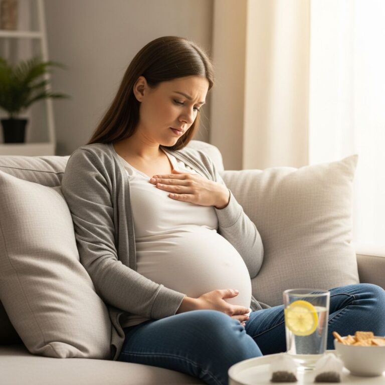
In women, oxytocin stimulates smooth muscle contractions in the myometrium (the muscular layer of the uterus) during labor, facilitating childbirth. Additionally, oxytocin triggers the milk-ejection reflex during lactation. When a nursing infant stimulates the nipple, sensory impulses travel through thoracic nerves to the hypothalamus, triggering the release of oxytocin from the posterior pituitary. The oxytocin then binds to receptors on myoepithelial cells surrounding the alveoli and small ducts in the breast, causing contractions that result in milk ejection.
Oxytocin also plays a role in the Ferguson reflex during labor. Cervical and vaginal stimulation triggers neural impulses that travel to the hypothalamus, prompting oxytocin release, which causes uterine contractions that facilitate delivery. At the cellular level, oxytocin stimulates smooth muscle contraction by increasing intracellular calcium concentration through voltage-dependent calcium channels, activating the contractile machinery of muscle cells.
How the Posterior Pituitary Works: The Hypothalamic-Hypophyseal System
The posterior pituitary operates as part of an integrated neuroendocrine system with the hypothalamus. Unlike the anterior pituitary, which is controlled by releasing hormones delivered through the hypophyseal portal blood system, the posterior pituitary is controlled through direct neural connections.
Hormone Synthesis and Transport: The hypothalamus synthesizes oxytocin and ADH in specialized neurosecretory neurons. These hormones are packaged together with their associated neurophysins (binding proteins) into secretory granules within the neurosecretory cells. These granules are then transported down the axons of the magnocellular neurons at a rate of 1-3 millimeters per hour, traveling through the infundibular stalk to reach terminals in the posterior pituitary.
Storage and Release: Once the neurosecretory granules reach the posterior pituitary, they accumulate in the Herring bodies at the nerve terminals. When the hypothalamus receives appropriate signals from the brain and body, it sends action potentials down these neurons. This electrical activity triggers the release of the hormones into the fenestrated sinusoidal capillaries surrounding the posterior pituitary. The hormones then enter the systemic circulation, where they travel throughout the body to reach their target organs.
This direct neural control mechanism is fundamentally different from the anterior pituitary’s hormonal control system. It allows for rapid, precise control of posterior pituitary hormone release in response to immediate physiological needs.
Functions and Target Organs
The hormones released by the posterior pituitary have widespread effects throughout the body:
Vasopressin (ADH): Primarily targets the kidneys to regulate water reabsorption and urine concentration, maintaining fluid balance and blood osmolality.
Oxytocin: Targets reproductive organs (uterus and mammary glands in women, vas deferens in men) to facilitate labor, milk ejection, and reproductive processes.
Common Posterior Pituitary Disorders
The main clinical issues related to the posterior pituitary involve abnormal hormone levels:
Hypersecretion: Excessive production and release of oxytocin or ADH can lead to complications such as inappropriate ADH syndrome (SIADH), characterized by water retention and hyponatremia (low blood sodium).
Hyposecretion: Insufficient production or release of these hormones can result in central diabetes insipidus (inadequate ADH) or reproductive dysfunction.
Development of the Posterior Pituitary
The posterior pituitary develops differently from the anterior pituitary. The neurohypophysis (posterior pituitary) develops from neuroectodermal tissue called the infundibulum, which grows inferiorly from the floor of the diencephalon (the region containing the thalamus and hypothalamus). Around 35 to 40 days of gestation, Rathke’s pouch (the embryonic precursor of the anterior pituitary) transforms into a vesicle that surrounds the anterolateral surface of the infundibulum before eventually detaching. The proliferation of the infundibulum diverticulum ultimately generates the cells that comprise the neurohypophysis.
Frequently Asked Questions
Q: What is the main difference between the posterior and anterior pituitary?
A: The posterior pituitary stores and releases two hormones (oxytocin and ADH) produced by the hypothalamus, while the anterior pituitary produces its own six hormones in response to releasing hormones from the hypothalamus. Additionally, the posterior pituitary is controlled by direct neural signals, whereas the anterior pituitary is controlled by hormonal signals.
Q: Why doesn’t the posterior pituitary produce its own hormones?
A: The posterior pituitary is functionally an extension of the hypothalamus and consists of neuronal tissue rather than glandular tissue. The hypothalamus produces the hormones and sends them down neural pathways to the posterior pituitary for storage and release.
Q: How does the body know when to release these hormones?
A: The hypothalamus receives sensory input and signals from the body. For ADH, osmoreceptors and volume receptors provide information about blood osmolality and volume. For oxytocin, signals from nursing stimulation or labor contractions trigger release through neural reflexes.
Q: What happens if the posterior pituitary is damaged?
A: Damage to the posterior pituitary can result in central diabetes insipidus (due to ADH deficiency) or problems with labor and lactation (due to oxytocin deficiency). The specific symptoms depend on which hormones are affected and the extent of the damage.
Q: Can medications affect posterior pituitary function?
A: Yes, certain medications can affect posterior pituitary hormone release. For example, some antipsychotic medications can increase prolactin levels and interfere with normal ADH regulation. Always discuss potential medication effects with your healthcare provider.
Q: Is the posterior pituitary the same size throughout life?
A: The posterior pituitary remains relatively stable in size throughout life, though hormonal production and responsiveness may change with age and physiological conditions such as pregnancy and lactation.
References
- Posterior pituitary — Wikipedia. Accessed December 2025. https://en.wikipedia.org/wiki/Posterior_pituitary
- The Posterior Pituitary Pathway — Global Library of Women’s Medicine. Accessed December 2025. https://www.glowm.com/section-view/heading/the-posterior-pituitary-pathway/item/283
- Physiology, Posterior Pituitary – StatPearls — National Center for Biotechnology Information (NCBI). 2023. https://www.ncbi.nlm.nih.gov/books/NBK526130/
- Posterior Pituitary: What It Is & Function — Cleveland Clinic. Accessed December 2025. https://my.clevelandclinic.org/health/body/23150-posterior-pituitary
- Neurohypophysis: Anatomy and function — Kenhub. Accessed December 2025. https://www.kenhub.com/en/library/anatomy/neurohypophysis
- The Pituitary Gland – Structure — TeachMeAnatomy. Accessed December 2025. https://teachmeanatomy.info/neuroanatomy/structures/pituitary-gland/
- Posterior Pituitary Hormones: What Are They — Osmosis. Accessed December 2025. https://www.osmosis.org/answers/posterior-pituitary-hormones
Similar Articles


Read full bio of medha deb