Pressure Ulcer Essential Guide: Causes, Prevention & Treatment
Comprehensive guide to pressure ulcers: causes, stages, prevention, and management strategies for at-risk patients.

A
pressure ulcer
, also known as apressure sore
,decubitus ulcer
, orbed sore
, is localized damage to the skin and underlying soft tissue usually over a bony prominence, resulting from prolonged pressure or pressure in combination with shear. These injuries commonly affect individuals with reduced mobility, such as the elderly, those bedridden, or wheelchair users, and can range from superficial skin changes to deep tissue destruction extending to muscle and bone.What is a Pressure Ulcer?
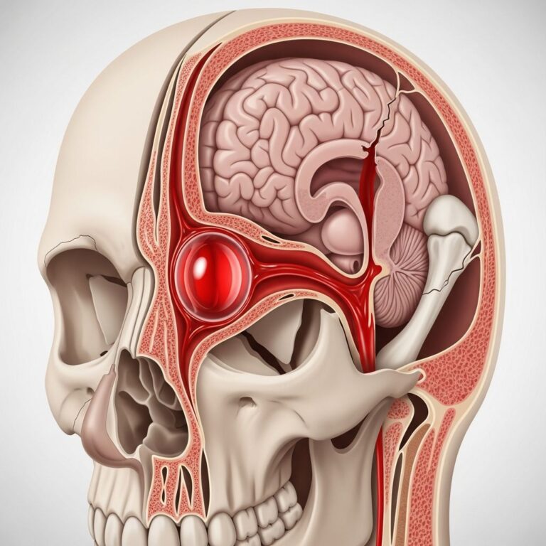
Pressure ulcers develop when sustained pressure on the skin impairs blood flow, leading to ischemia and tissue necrosis. This typically occurs over bony prominences like the sacrum, heels, hips, elbows, and shoulders where body weight concentrates force. The condition was formerly called decubitus ulcers, emphasizing the ‘lying down’ position, but ‘pressure ulcer’ better reflects multifactorial causes including shear (sliding forces) and friction. According to refined definitions, it is “an area of localized soft tissue ischemic necrosis caused by prolonged pressure higher than capillary pressure (16-33 mm Hg) with or without shear, related to posture, usually over a bony prominence”.
Muscles and subcutaneous tissues are more vulnerable than skin, so visible surface damage often underrepresents deeper injury—the ‘tip of the iceberg’ phenomenon. Ulcers can form rapidly, within 2 hours of unrelieved pressure, due to occluded capillaries starving tissues of oxygen and nutrients.
Who is at Risk of Pressure Ulcers?
Anyone can develop pressure ulcers, but risk escalates with immobility, where body weight continuously presses on the same skin areas. Key risk groups include:
- Older adults with frailty or limited mobility.
- Patients with spinal cord injuries, strokes, or neuropathies impairing sensation and movement.
- Wheelchair-bound individuals (e.g., ischial tuberosity pressure from sitting).
- Critically ill or hospitalized patients.
- Those with incontinence, poor nutrition, edema, or vascular diseases reducing tissue perfusion.
Neurological deficits prevent pain perception, delaying posture changes, while edema compromises circulation despite seeming cushioning. Tools like the Braden Scale assess risk based on mobility, activity, sensory perception, moisture, nutrition, and friction/shear.
What are the Causes of Pressure Ulcers?
Primary cause is
prolonged pressure
exceeding capillary closing pressure, occluding blood vessels and causing anoxia. An inverse relationship exists: higher pressure acts faster than lower sustained pressure.Contributing Factors
- Shear: Gravity pulls body down while fixed upper body (e.g., against bed headboard) stretches tissues, kinking vessels.
- Friction: Rubbing skin against surfaces during dragging.
- Moisture: From incontinence or sweat macerates skin, increasing fragility.
- Positioning: Supine (sacrum), prone (knees/toes), lateral (trochanter), sitting (ischium).
Post-relief reperfusion triggers inflammation, exacerbating damage via macrophages.
What are the Signs and Symptoms of Pressure Ulcers?
Early signs include non-blanchable
discoloration
(red on light skin, purple/blue on darker), warmth, swelling, induration, pain, or itchiness. Skin may feel boggy or spongy before breaking. Advanced symptoms involve blisters, open wounds, or necrotic tissue.| Early Signs | Advanced Signs |
|---|---|
| Persistent erythema, warmth, edema | Open wounds, blisters, slough/eschar |
| Pain/tenderness, sponginess | Foul odor, exudate, infection signs |
How are Pressure Ulcers Staged?
The National Pressure Injury Advisory Panel (NPIAP) stages ulcers by tissue damage depth.
Stage 1
Intact skin with non-blanchable redness (purple on darker skin). Warm, cool, firm, or boggy.
Stage 2
Partial-thickness loss of dermis: shallow open wound or intact/ruptured blister with pink/red bed, no slough.
Stage 3
Full-thickness loss: subcutaneous fat visible, but not muscle/bone. Slough may obscure.
Stage 4
Full-thickness loss exposing muscle, tendon, bone. Undermining/tunneling, eschar/slough.
Unstageable
Full-thickness with base obscured by slough/eschar.
Deep Tissue Pressure Injury (DTPI)
Purple/maroon intact skin or blood-filled blister indicating deeper necrosis.
Complications of Pressure Ulcers
Untreated ulcers lead to cellulitis, osteomyelitis, sepsis, or even death. Stage 2+ risk infection; deep ulcers expose bone to bacteria.
How is a Pressure Ulcer Diagnosed?
Diagnosis relies on visual inspection, staging, and history of risk factors. Wound measurement (length, width, depth) tracks progress. Swabs rule out infection; imaging (X-ray/MRI) detects osteomyelitis.
What is the Treatment for Pressure Ulcers?
Treatment addresses pressure relief, wound healing, and complications.
General Management
- Pressure redistribution: Special mattresses/overlays, frequent repositioning (q2h).
- Cleaning: Normal saline, no hydrogen peroxide.
- Dressings: Foams, hydrocolloids for stage 2; match exudate.
Stage-Specific
| Stage | Treatment |
|---|---|
| 1-2 | Protect, moisture barrier, topical agents |
| 3-4 | Debridement (enzymatic/sharp), negative pressure therapy |
| Unstageable/DTPI | Debride after eschar assessment, surgical consult |
Advanced
Surgery (flaps, grafts) for non-healing stage 4; antibiotics for infection. Nutrition: High protein, vitamins A/C, zinc.
Prevention of Pressure Ulcers
Prevention outperforms treatment.
- Repositioning: Every 2 hours; use 30-degree lateral positioning.
- Support Surfaces: Air-fluidized beds, low-air-loss mattresses.
- Skin Care: Clean/dry, barrier creams for moisture.
- Nutrition: Adequate calories/protein; screen at-risk.
- Mobility: Encourage movement, heel elevation.
Staff education via CNA training is vital.
Pressure Ulcer in Specific Populations
Wheelchair Users
Ischial pressure from sitting; weight shifts q15-30min.
Paediatric/Neonates
Occipital pressure in infants; adjust for growth.
Frequently Asked Questions
What is the most common site for pressure ulcers?
Sacrum in bedbound; heels/ischia in seated.
Can pressure ulcers heal?
Yes, stages 1-2 heal with care; stage 4 may require surgery.
How quickly do pressure ulcers form?
As fast as 2 hours in high-risk.
Is nutrition important?
Critical; malnutrition delays healing.
What role does incontinence play?
Moisture causes maceration, raising risk.
References
- Stage 2 Pressure Ulcers: Symptoms, Causes, and Solutions — Quality Insights. 2023. https://www.qualityinsights.org/nursing-home-insights/stage-2-pressure-ulcers
- Pressure Ulcers: Back to the Basics — PMC / PubMed Central. 2012-10-31. https://pmc.ncbi.nlm.nih.gov/articles/PMC3495374/
- Sepsis and Pressure Ulcers (Pressure Injuries) — Sepsis Alliance. 2024. https://www.sepsis.org/sepsisand/pressure-ulcers-pressure-injuries/
- Pressure Ulcers (Pressure Sores) — NHS. 2023. https://www.nhs.uk/conditions/pressure-sores/
- Pressure Ulcers and Wounds/Injury Management — AAPM&R. 2024. https://www.aapmr.org/about-physiatry/conditions-treatments/medical-rehabilitation/pressure-ulcers-and-wounds
- Bedsores (Pressure Ulcers): Symptoms, Staging & Treatment — Cleveland Clinic. 2023-07-28. https://my.clevelandclinic.org/health/diseases/17823-bedsores-pressure-injuries
- Bedsores (Pressure Ulcers) – Symptoms and Causes — Mayo Clinic. 2023-08-01. https://www.mayoclinic.org/diseases-conditions/bed-sores/symptoms-causes/syc-20355893
Similar Articles


Read full bio of medha deb