Pretibial Myxedema: Causes, Symptoms & Treatment
Understanding pretibial myxedema: An autoimmune skin condition linked to Graves' disease.

What Is Pretibial Myxedema?
Pretibial myxedema, also known as localized myxedema or thyroid dermopathy, is a skin condition characterized by the accumulation of excess glycosaminoglycans in the dermis and subcutaneous tissue of the skin. The primary glycosaminoglycan involved in this condition is hyaluronic acid, a complex carbohydrate that plays a crucial role in tissue hydration and lubrication. These substances are produced by specialized cells called fibroblasts, which become overactive in response to autoimmune processes.
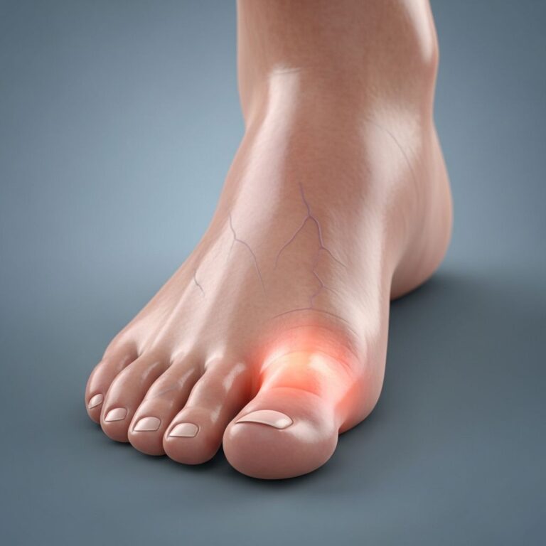
This condition represents an autoimmune manifestation most commonly associated with Graves’ disease, an autoimmune disorder affecting the thyroid gland. While pretibial myxedema can occasionally occur in Hashimoto’s thyroiditis, it is predominantly linked to Graves’ disease and its associated complications. The condition typically affects the skin on the front of the lower leg, just above the ankle, though it can occasionally appear in other areas of the body, indicating a more systemic process.
In most cases, pretibial myxedema is asymptomatic and presents primarily as a cosmetic concern rather than a symptomatic problem. However, in more severe manifestations, the condition can progress to elephantiasis, a significant swelling and thickening of the affected tissue, or thyroid acropachy, which involves thickening of the skin and soft tissues in extremities.
Causes and Risk Factors
Pretibial myxedema develops as a result of multiple interconnected factors working in conjunction. The primary cause is the autoimmune response characteristic of Graves’ disease, where thyroid-stimulating hormone receptor (TSH-R) antibodies attack the thyroid gland, causing hyperthyroidism. These same antibodies can cross-react with TSH receptors located in the connective tissue of the skin, initiating a similar autoimmune cascade.
The development of pretibial myxedema involves both humoral and cellular immune mechanisms. Thyroid-stimulating hormone receptors in the connective tissue serve as the antigen responsible for triggering the immune process, similar to what occurs in Graves’ ophthalmopathy. Once activated, fibroblasts produce excessive amounts of glycosaminoglycans, leading to the characteristic accumulation of mucin in the skin layers.
The localization of pretibial myxedema predominantly in the shin area relates to both mechanical and positional factors. The dependent position of the legs combined with the mechanical stress placed on the pretibial region during daily activities appears to create an environment favorable for glycosaminoglycan accumulation. Additionally, the pretibial area has specific lymphatic drainage patterns and tissue characteristics that make it particularly susceptible to this type of mucinous deposition.
Several demographic and clinical factors increase the risk of developing pretibial myxedema. Patients with severe Graves’ disease, particularly those with significant ophthalmopathy, face a higher risk. The presence of high serum concentrations of thyroid-stimulating hormone receptor antibodies indicates both the severity of the autoimmune condition and the likelihood of developing dermopathy. Almost all cases of thyroid dermopathy are associated with relatively severe ophthalmopathy, and typically, ophthalmopathy appears first while dermopathy develops much later.
Symptoms and Clinical Presentation
The clinical presentation of pretibial myxedema varies considerably depending on disease severity. In mild to moderate cases, the condition is often completely asymptomatic, with patients noticing only cosmetic changes to their skin. The affected skin typically appears thickened, with a characteristic non-pitting edema that does not leave an indentation when pressure is applied.
The most common manifestation is nonpitting edema, which accounts for approximately 43.3% of cases. The pretibial area, or the front of the lower leg above the ankle, is the region most commonly involved, occurring in about 99.4% of cases. The skin may appear slightly raised, with a waxy or shiny appearance, and may have a slightly reddish or flesh-colored hue.
In more severe cases, patients may experience:
- Persistent swelling and thickening of the affected skin
- Discomfort or mild pain in the affected area
- Difficulty fitting into shoes or wearing tight-fitting clothing
- Cosmetic concerns affecting quality of life and self-confidence
- Progression to elephantiasis in advanced cases, characterized by significant tissue enlargement
Advanced forms of dermopathy can lead to elephantiasis, where the affected tissue becomes markedly enlarged and thickened, resembling the appearance associated with parasitic infections. Additionally, thyroid acropachy may develop, affecting the fingers and toes with similar thickening and skin changes.
Connection to Graves’ Disease and Other Thyroid Conditions
Pretibial myxedema is almost exclusively associated with Graves’ disease, an autoimmune thyroid disorder characterized by hyperthyroidism. Graves’ disease causes thyroid-stimulating hormone receptor antibodies to activate these receptors on thyroid cells, leading to excessive production of thyroid hormones and the classic symptoms of hyperthyroidism, including weight loss, palpitations, sweating, and tremors.
The relationship between Graves’ disease and pretibial myxedema is so consistent that the presence of dermopathy in a patient with hyperthyroidism typically confirms the diagnosis of Graves’ disease rather than other causes of hyperthyroidism. Among patients with Graves’ disease and dermopathy, the vast majority—approximately 97%—also have ophthalmopathy, an eye condition involving inflammation, exophthalmos (eye protrusion), and potential vision complications.
While pretibial myxedema occasionally occurs in Hashimoto’s thyroiditis, another autoimmune thyroid condition, this is relatively rare. Hashimoto’s thyroiditis typically causes hypothyroidism rather than hyperthyroidism and is more commonly associated with other skin manifestations. It is important to distinguish pretibial myxedema from myxedema coma, a severe complication of poorly controlled hypothyroidism that represents a medical emergency.
Diagnosis and Diagnostic Tests
The diagnosis of pretibial myxedema is based on a combination of clinical examination, patient history, and laboratory findings. A healthcare provider will typically begin with a thorough physical examination, looking for characteristic skin changes on the pretibial region or other areas of the body.
Key diagnostic components include:
- Clinical History: Documentation of a history of Graves’ hyperthyroidism and associated ophthalmopathy
- Physical Examination: Assessment of typical pretibial skin lesions showing nonpitting edema and characteristic appearance
- Laboratory Testing: Measurement of thyroid function including TSH levels and serum TSH receptor antibodies
- Skin Biopsy: In some cases, skin biopsy may be performed to confirm the diagnosis
Laboratory tests typically reveal low serum TSH concentrations and elevated thyroid hormone levels consistent with hyperthyroidism. Importantly, all patients with localized myxedema have high serum concentrations of thyroid-stimulating hormone receptor antibodies, reflecting the severity of the underlying autoimmune condition.
When a skin biopsy is performed, histopathological examination typically reveals deposition of mucin (glycosaminoglycans) throughout the dermis and subcutaneous tissue. This histological finding confirms the diagnosis, though many clinical cases can be diagnosed based on characteristic presentation without requiring biopsy.
Treatment Options
Conservative Management
For many patients with pretibial myxedema, conservative management and observation may be the appropriate approach. Since the majority of cases are asymptomatic and cause only cosmetic concerns, many patients do not require active therapy. Minimization of risk factors, including proper thyroid hormone control and management of underlying Graves’ disease, is fundamental to all treatment approaches.
Topical Corticosteroid Therapy
Topical corticosteroids represent the most commonly used treatment modality, employed in approximately 53.9% of cases. These medications are applied directly to the affected skin, often under occlusive dressing to enhance penetration and efficacy. In mildly severe symptomatic cases with cosmetic concerns, topical corticosteroids applied under occlusive dressing have demonstrated beneficial effects.
Topical corticosteroids are offered in varying potencies, from low-potency options including fluocinolone acetonide to high-potency formulations such as clobetasole propionate. The choice of corticosteroid potency depends on disease severity and individual patient factors. Regular application, often daily or multiple times daily, is typically required for optimal results.
Systemic Therapies
In more severe cases, systemic immunomodulation may be necessary, though conclusive evidence for long-term efficacy of these modalities remains limited. Systemic treatments may include oral or intravenous glucocorticoids, which have shown success in some case reports. However, the decision to use systemic therapy must be carefully weighed against potential adverse effects and the chronic nature of the condition.
Additional systemic and adjunctive treatments reported to show varying degrees of success in small studies include:
- Intravenous immunoglobulin
- Octreotide
- Pentoxifylline
- Plasmapheresis
- Radiotherapy
- Intralesional steroids
Supportive and Compressive Therapies
When significant edema and elephantiasis are present, local compressive therapy may provide additional benefit. Compression garments and dressings help reduce fluid retention in the affected tissue and may improve skin appearance and comfort. These conservative approaches are often combined with topical corticosteroid therapy for enhanced efficacy.
Surgical Considerations
Surgical excision has been attempted in some cases but is generally not advised due to high rates of dermopathy recurrence. Despite success in some individual case studies, the recurrent nature of the condition makes surgical intervention typically ineffective as a definitive treatment. Surgical options are generally reserved for severe cases that have failed other interventions.
Prognosis and Long-Term Outcomes
The prognosis for pretibial myxedema is generally favorable, particularly for milder cases. The likelihood of remission depends significantly on the severity of the initial disease rather than on the specific treatment received. Long-term follow-up studies have provided valuable insights into the natural history of this condition.
Studies show that after 25 years of follow-up, approximately 70% of milder untreated cases experienced partial or complete remission, compared to 58% of severe cases treated with local therapy. The myxedema clears completely in the majority of patients with mild disease, while even more severe disease resolves in more than half of patients after several years.
The elephantiasic form of the disease represents the most challenging presentation, being the most difficult to treat and least likely to clear completely. Importantly, severe cases that receive topical corticosteroids or other therapies do not demonstrate significantly better outcomes than untreated milder cases, suggesting that natural disease evolution plays a major role in determining prognosis.
Most patients with asymptomatic pretibial myxedema do not require ongoing treatment or intensive follow-up once the diagnosis is established. Management typically focuses on adequate control of the underlying Graves’ disease and thyroid dysfunction, which may indirectly benefit the skin manifestation.
When to Seek Medical Attention
Patients with Graves’ disease or known hyperthyroidism should inform their healthcare provider about any skin changes affecting the lower legs or other areas of the body. A dermatology referral may be appropriate when cosmetic concerns warrant discussion of treatment options or when the diagnosis is unclear. Patients should seek medical attention if:
- Skin lesions appear to progress rapidly or become painful
- Swelling interferes with daily activities or causes functional impairment
- Cosmetic concerns significantly affect quality of life
- New skin manifestations develop beyond the pretibial area
- The condition fails to improve with standard management approaches
Frequently Asked Questions
Q: Is pretibial myxedema dangerous or life-threatening?
A: Pretibial myxedema itself is not dangerous or life-threatening. The condition is primarily a cosmetic concern and usually asymptomatic. However, the underlying Graves’ disease requires proper treatment to prevent complications such as thyroid storm or cardiac arrhythmias.
Q: Can pretibial myxedema disappear on its own?
A: Yes, pretibial myxedema can remit spontaneously, particularly in mild cases. Approximately 70% of milder untreated cases achieve partial or complete remission within several years. Even severe cases show remission in more than half of patients after extended periods.
Q: Does treating Graves’ disease help resolve pretibial myxedema?
A: While proper management of Graves’ disease is important for overall health, it does not necessarily prevent or resolve pretibial myxedema. However, adequate thyroid control forms the foundation of all management strategies and may provide indirect benefits to skin manifestations.
Q: What is the difference between pretibial myxedema and myxedema coma?
A: Pretibial myxedema is a skin condition associated with Graves’ disease (hyperthyroidism), while myxedema coma is a severe, life-threatening complication of severe hypothyroidism. These are distinct conditions affecting different thyroid function states.
Q: Are there any side effects from topical corticosteroid treatment?
A: Long-term topical corticosteroid use can cause skin atrophy, striae (stretch marks), and other local effects. Regular monitoring by a healthcare provider helps minimize these risks while maximizing therapeutic benefits.
Q: Can pretibial myxedema affect other areas besides the shins?
A: While the pretibial area is involved in approximately 99.4% of cases, pretibial myxedema can occasionally occur in other areas of the body, indicating a more systemic autoimmune process. When the condition extends beyond the pretibial region, it suggests a broader manifestation of the underlying autoimmune thyroid disease.
References
- Pretibial myxedema: pathophysiology and treatment options — National Center for Biotechnology Information. 2004-11-01. https://pubmed.ncbi.nlm.nih.gov/16252929/
- Pretibial myxedema in Grave’s disease: A case report and treatment — National Institutes of Health. 2024. https://pmc.ncbi.nlm.nih.gov/articles/PMC10883341/
- Pretibial myxoedema — DermNet New Zealand. 2024. https://dermnetnz.org/topics/pretibial-myxoedema
- Dermopathy of Graves’ Disease (Pretibial Myxedema): Long-Term Outcome — Journal of Clinical Endocrinology & Metabolism. 2002. https://academic.oup.com/jcem/article/87/2/438/2846476
- Myxedema: Symptoms, treatment & coma — Medical News Today. 2024. https://www.medicalnewstoday.com/articles/321886
Similar Articles


Read full bio of medha deb