Primary Congenital Glaucoma: Diagnosis and Care
Explore the essential strategies for identifying and managing primary congenital glaucoma in infants to safeguard their vision.

Primary congenital glaucoma (PCG) represents a pressing challenge in pediatric ophthalmology, emerging at birth due to developmental flaws in the eye’s drainage system. This condition, responsible for a notable share of childhood blindness cases globally, demands swift recognition and intervention to preserve vision. Unlike typical adult glaucoma, PCG stems from isolated trabecular meshwork anomalies without associated systemic issues, affecting roughly 1 in 10,000 newborns. Early action can yield success rates exceeding 80%, highlighting the critical window for treatment.
Understanding the Roots of PCG
The core issue in PCG lies in the trabecular meshwork, the eye’s natural filter for aqueous humor. Genetic mutations, particularly in genes like CYP1B1, disrupt this structure, causing fluid buildup and elevated intraocular pressure (IOP). This pressure stretches the compliant infant eye, leading to characteristic changes. While no preventive measures exist today, genetic screening in high-risk families—such as those with consanguinity in regions like Saudi Arabia or Slovakia—offers future promise for proactive care.
Spotting the Warning Signs in Infants
Parents and caregivers often notice initial clues.
Buphthalmos
, or eye enlargement, occurs as high IOP expands the sclera.Corneal clouding
arises from edema, sometimes accompanied byHaab striae
—horizontal breaks in Descemet’s membrane visible under slit-lamp illumination. Infants may exhibit excessive tearing, light sensitivity (photophobia), and blepharospasm, mimicking conjunctivitis but persisting. These signs typically surface between 3-6 months, though diagnosis can extend to age 3. Unchecked, they progress to optic nerve damage and potential blindness.- Enlarged eye globe (buphthalmos)
- Cloudy or hazy cornea
- Haab striae on corneal exam
- Profuse tearing and photophobia
- Eye rubbing or spasms
Comprehensive Diagnostic Approach
Confirming PCG requires exams under anesthesia due to infants’ non-cooperation. Key steps include IOP measurement via tonometry, which reveals pressures often above 21 mmHg, alongside corneal diameter assessment (normal <11 mm at birth, <12 mm by 1 year). Gonioscopy unveils a wide-angle with iris insertion into the trabecular meshwork, distinguishing PCG from secondary forms like Axenfeld-Rieger syndrome. Axial length via A-scan ultrasound detects elongation beyond age norms, tracked every 3-4 months.
Post-dilation, retinoscopy identifies refractive errors like myopia from corneal distortion. Fundus evaluation spots optic disc cupping, which may reverse with IOP control. Anterior segment OCT in older treated cases shows endothelial changes. Diagnosis hinges on elevated IOP with ocular enlargement sans other anomalies.
| Diagnostic Tool | Purpose | Typical Findings in PCG |
|---|---|---|
| Tonometry | Measure IOP | >21 mmHg, often much higher |
| Gonioscopy | View angle structures | High iris insertion, no scleral spur |
| Slit-lamp exam | Assess cornea | Haab striae, edema |
| A-scan ultrasound | Measure axial length | Elongated beyond age-matched norms |
| Fundoscopy | Evaluate optic nerve | Increased cup-to-disc ratio |
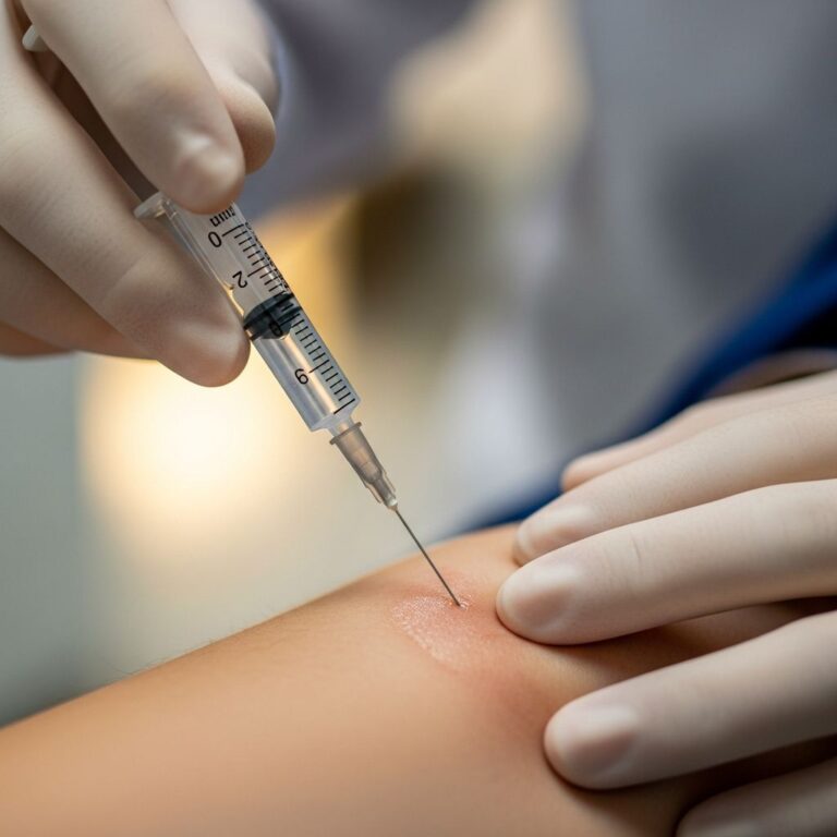
Surgical Interventions: The Frontline Defense
Surgery stands as the cornerstone of PCG management, bypassing medications’ limited efficacy in infants. Angle procedures like
goniotomy
andtrabeculotomy
boast 80-90% initial success by incising the trabecular meshwork to enhance outflow. Goniotomy uses a blade through a corneal incision to open the inner meshwork, ideal for clear corneas. Trabeculotomy, accessing externally or via 360° circumferential cleavage, suits opaque corneas.Many infants require multiple surgeries; combined 360° approaches yield superior outcomes. If angle surgery falters, filtering procedures like trabeculectomy or tube shunts create alternative drainage paths. Laser options, such as cyclophotocoagulation, target ciliary body fluid production as salvage therapy. Timing is crucial—bilateral cases often undergo simultaneous operations to minimize anesthesia risks.
Medical Support and Adjunctive Therapies
While surgery predominates, topical agents bridge gaps.
Carbonic anhydrase inhibitors
(e.g., dorzolamide) reduce aqueous production safely as first-line. Prostaglandin analogs and beta-blockers follow, though long-term pediatric data is sparse. Oral agents or drops control IOP pre-surgery. Post-op, medications sustain pressure control, with vigilant monitoring for amblyopia in unilateral cases.Navigating Long-Term Management and Prognosis
Success hinges on ongoing surveillance. Regular exams track IOP, corneal clarity, and optic nerve health, adjusting for growth-related changes. About 50% need additional surgeries, but controlled IOP often yields 20/60 or better vision in the better eye. Risks include corneal scarring, amblyopia, and persistent cupping. Multidisciplinary care involving pediatricians aids holistic development.
Prognosis shines with early intervention: 80-90% achieve stable vision. Variable outcomes stem from mutation severity and promptness of care. Families in high-prevalence areas benefit from awareness campaigns.
FAQs on Primary Congenital Glaucoma
What causes primary congenital glaucoma?
PCG arises from genetic defects in the eye’s drainage angle, leading to fluid buildup and high pressure.
At what age is PCG typically diagnosed?
Signs appear by 3-6 months, with diagnosis possible up to age 3.
Is surgery always necessary for PCG?
Yes, angle surgeries like goniotomy or trabeculotomy are primary treatments, with medications as adjuncts.
Can PCG be prevented?
No current prevention exists, but genetic screening in at-risk families may enable early detection.
What is the long-term outlook for children with PCG?
Early treatment offers 80-90% success, potentially preserving good vision.
References
- Primary Congenital Glaucoma – EyeWiki — American Academy of Ophthalmology EyeWiki. 2023. https://eyewiki.org/Primary_Congenital_Glaucoma
- Glaucoma in Children — American Association for Pediatric Ophthalmology and Strabismus. 2023. https://aapos.org/glossary/glaucoma-in-children
- Chapter 10. Pediatric Glaucoma – EyeRounds — University of Iowa. 2023. https://eyerounds.org/books/glaucoma_guide/chapter10.html
- Primary Congenital Glaucoma: Causes, Symptoms, and Treatment — WebMD. 2023. https://www.webmd.com/eye-health/primary-congenital-glaucoma
- Childhood Glaucoma: Diagnosis, Treatment, And Hope — Glaucoma Research Foundation. 2023. https://glaucoma.org/articles/childhood-glaucoma-diagnosis-treatment-and-hope
- Primary Congenital Glaucoma – StatPearls — NCBI Bookshelf. 2023-10-01. https://www.ncbi.nlm.nih.gov/books/NBK574553/
Similar Articles


Read full bio of medha deb