Primary Cutaneous Anaplastic Large Cell Lymphoma
Rare indolent skin lymphoma with CD30+ cells: symptoms, diagnosis, treatment, and excellent prognosis explored in depth.

Primary cutaneous anaplastic large-cell lymphoma (pcALCL) is a rare subtype of indolent cutaneous T-cell lymphoma (CTCL) defined by CD30-positive atypical large lymphocytes infiltrating the skin. It belongs to the spectrum of primary cutaneous CD30+ lymphoproliferative disorders, which also includes lymphomatoid papulosis, and typically remains confined to the skin without extracutaneous spread at diagnosis.
Introduction
pcALCL represents about 8-12% of all cutaneous T-cell lymphomas and is characterized by its indolent behaviour despite aggressive histological features. Unlike systemic anaplastic large-cell lymphoma (ALCL), pcALCL arises primarily in the skin and has a favourable prognosis, with 5-year survival rates exceeding 90% in localized cases. The disease manifests as solitary or multifocal nodules or tumours, often ulcerating, and spontaneous regression occurs in 20-40% of lesions. This article details demographics, causes, clinical presentation, diagnosis, treatment, and outcomes based on established medical literature.
Demographics
pcALCL affects individuals across all ages and races, but shows a marked male predominance (male-to-female ratio of 3:1). The peak incidence is between 50-70 years, though paediatric and even congenital cases are documented. It is uncommon in children under 10, comprising less than 1% of paediatric lymphomas, but adolescent cases mirror adult presentations. Globally, incidence is low at approximately 0.1-0.2 cases per 100,000 persons annually, with no strong geographic or ethnic biases reported.
Causes
The exact aetiopathogenesis of pcALCL remains unclear, with no definitive risk factors identified. It is an acquired condition, not hereditary or contagious, arising from clonal expansion of post-thymic T-cells in the skin. Hypotheses include:
- Disrupted CD30 signalling pathways promoting lymphoid cell survival and proliferation.
- Restricted T-cell repertoire in peripheral blood, suggesting immune dysregulation.
- Reactive phenomena, such as chronic antigen stimulation, leading to CD30 overexpression.
- Genetic alterations like monoclonal T-cell receptor gene rearrangements, but unlike systemic ALCL, ALK gene translocations are absent in over 90% of cases (ALK-negative).
No viral associations (e.g., HTLV-1, EBV) or environmental triggers are consistently linked, distinguishing it from other CTCLs like mycosis fungoides.
Clinical Features
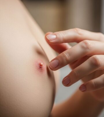
Patients typically present with solitary or localized nodules or tumours (60-80% of cases), progressing to multifocal disease in 20%. Lesions are raised, red to violaceous, often >2 cm, and prone to central ulceration or necrosis. Common sites include extremities (arms, legs), trunk, face, and buttocks; oral mucosa involvement is rare.
Associated symptoms may include:
- Pain, tenderness, or pruritus in 30-50% of cases.
- Regional lymphadenopathy in 10-20%, usually reactive rather than malignant.
- B symptoms (fever, weight loss, night sweats) are uncommon (<10%), unlike systemic ALCL.
In darker skin tones, lesions may appear hyperpigmented or hypopigmented. Paediatric cases often present as solitary limb nodules with excellent response to therapy.
Complications
Despite its indolent nature, complications arise from disease progression or treatment:
- Local recurrence in up to 50% of cases.
- Extracutaneous spread (lymph nodes, viscera) in 10-25%, portending poorer outcomes.
- Secondary bacterial infections from ulcerated lesions.
- Treatment-related effects: radiation-induced atrophy, surgical scarring, or chemotherapy toxicity.
- Rare transformation to aggressive systemic lymphoma.
Multifocal disease increases risk of lymph node involvement (10%).
Diagnosis
Diagnosis hinges on clinicopathological correlation, excluding systemic involvement. Key steps include:
- Skin biopsy: Shows diffuse infiltrates of large anaplastic cells (>75% CD30+ by immunohistochemistry). Hallmark ‘wreath-like’ nuclei and Reed-Sternberg-like cells are seen.
- Immunophenotyping: CD4+, CD30+, ALK-, EMA-, often cytotoxic markers (granzyme B+).
- T-cell receptor gene rearrangement: Clonal in most cases.
- Staging investigations: Blood tests (CBC, LDH), PET-CT or CT scans for nodes/viscera, bone marrow biopsy if indicated.
TNM staging: T1 (single lesion <5cm), T2 (multiple regional), T3 (multifocal/large >5cm). Specialist pathologist review is essential to differentiate from mimics.
Differential Diagnoses
| Condition | Key Distinguishing Features |
|---|---|
| Lymphomatoid papulosis (LyP) | Smaller papules (crops), self-resolve in weeks; shares CD30+ histology but polyclonal |
| Systemic ALCL | ALK+ in 50-80%, extracutaneous at onset; requires imaging to exclude |
| Transformed mycosis fungoides | Pre-existing plaques/patches; epidermotropism, CD30 variable |
| CD30+ B-cell lymphoproliferative disorders | B-cell markers (CD20+), less anaplastic |
| Infectious/reactive (e.g., arthropod bite) | Polyclonal, resolves quickly; negative clonality |
Treatment
Treatment is stage-dependent, prioritizing skin-directed therapies due to indolent course. First-line options:
- Solitary/regional lesions (T1/T2): Surgical excision (Mohs micrographic surgery ideal) or low-dose radiotherapy (e.g., 20-30 Gy); complete response >95%.
- Multifocal (T3): Radiotherapy, low-dose methotrexate (10-25 mg/week), or IFN-alpha.
For extracutaneous disease:
- Brenntuximab vedotin (anti-CD30 antibody-drug conjugate): High response rates (70-90%), now first-line systemic.
- Multi-agent chemotherapy (CHOP) reserved for refractory cases due to relapse risk.
- Phototherapy (e.g., PUVA) or topical chemotherapy (mechlorethamine) for superficial disease.
Up to 40% regress spontaneously; observation is viable for small lesions.
Outcome
pcALCL has an excellent prognosis: 5-year disease-specific survival 95% overall, dropping to 50-80% with extracutaneous spread. Recurrence occurs in 50%, mostly cutaneous; 25% of recurrences involve nodes.
Good prognostic factors:
- Limited skin involvement (T1/T2).
- Spontaneous regression.
- Absence of extracutaneous disease.
Poor prognostic factors:
- Multifocal/large tumours.
- Lymph node involvement.
- Ulceration.
Long-term surveillance (every 3-6 months) is recommended.
Frequently Asked Questions (FAQs)
What is primary cutaneous anaplastic large-cell lymphoma?
A rare, slow-growing skin cancer of T-cells marked by CD30+ large cells, usually staying in the skin.
Who gets pcALCL?
Mostly men aged 50-70, but all ages/races possible.
Does it spread beyond the skin?
Rarely (10-25%); mostly local nodes.
How is it treated?
Surgery or radiation for localized; systemic agents like brentuximab for advanced.
What is the prognosis?
Excellent: 95% 5-year survival.
Is it related to lymphomatoid papulosis?
Yes, both CD30+ cutaneous disorders, but LyP is papular and self-resolving.
References
- Primary cutaneous anaplastic large-cell lymphoma — DermNet NZ. 2023. https://dermnetnz.org/topics/primary-cutaneous-anaplastic-large-cell-lymphoma
- Primary Cutaneous Anaplastic Large Cell Lymphoma — MalaCards. 2024. https://www.malacards.org/card/primary_cutaneous_anaplastic_large_cell_lymphoma
- Anaplastic Large Cell Lymphoma: Symptoms, Causes & Treatment — Cleveland Clinic. 2024-01-15. https://my.clevelandclinic.org/health/diseases/24029-anaplastic-large-cell-lymphoma
- Subtype Fact Sheet: Primary Cutaneous Anaplastic Large Cell Lymphoma — Lymphoma Coalition. 2022. https://lymphomacoalition.org/wp-content/uploads/LC_CutaneousLymphoma_VF_A4_SubtypeFactSheet_PCALCL.pdf
Similar Articles


Read full bio of Sneha Tete


