Primary Cutaneous Follicle Centre Lymphoma
Comprehensive guide to primary cutaneous follicle centre lymphoma: symptoms, diagnosis, treatment, and prognosis for this indolent skin lymphoma.

Authoritative facts about primary cutaneous follicle centre lymphoma (PCFCL), the most common type of primary cutaneous B-cell lymphoma affecting the skin.
What is primary cutaneous follicle centre lymphoma?
Primary cutaneous follicle centre lymphoma (PCFCL) represents the most prevalent form of cutaneous B-cell lymphoma, accounting for over half of all cases. It is classified as an indolent, low-grade non-Hodgkin lymphoma originating from follicle centre cells, specifically centrocytes and centroblasts, within the skin without evidence of extracutaneous involvement at diagnosis. Unlike its systemic counterpart, PCFCL has distinct genetic and clinical features, including rare t(14;18) translocations and frequent mutations in chromatin-modifying genes.
PCFCL typically affects middle-aged to older adults, with a median age of presentation around 60 years, showing a slight male predominance. It arises de novo in the skin and is listed in the World Health Organization-European Organization for Research and Treatment of Cancer (WHO-EORTC) classification as a distinct entity due to its favourable prognosis and unique histopathological profile. The disease remains confined to the skin in most cases, with regional lymph node involvement being uncommon (less than 10%).
Histologically, PCFCL is characterized by neoplastic follicles or diffuse infiltrates of small cleaved centrocytes (resembling germinal centre cells) and variable centroblasts, often separated from the epidermis by a Grenz zone. The neoplastic cells express B-cell markers such as CD19, CD20, CD79a, and follicle centre markers like CD10 and BCL-6, while typically lacking BCL-2 expression and showing low Ki-67 proliferation index.
Who gets primary cutaneous follicle centre lymphoma?
Primary cutaneous follicle centre lymphoma predominantly affects adults over 50 years of age, with a peak incidence in the sixth to seventh decades of life. Men are affected slightly more often than women, with a male-to-female ratio of approximately 1.5:1. There is no strong association with specific risk factors such as UV exposure, immunosuppression, or viral infections, distinguishing it from other cutaneous lymphomas.
- Age: Median 60 years (range 30–90 years)
- Sex: Male predominance
- Risk factors: None definitively identified; rare familial cases reported
PCFCL is uncommon in children and young adults. It can occur in patients with a history of other lymphoproliferative disorders, but primary presentation in otherwise healthy individuals is typical.
What causes primary cutaneous follicle centre lymphoma?
The precise aetiology of PCFCL remains unknown, but molecular studies reveal somatic mutations in genes involved in chromatin remodelling (e.g., CREBBP, KMT2D), epigenetic modification, and B-cell signalling pathways. Unlike systemic follicular lymphoma, PCFCL rarely harbours the IGH-BCL2 translocation t(14;18), which occurs in less than 10% of cases. Aberrant somatic hypermutation targeting immunoglobulin variable genes and histone-modifying enzymes drives lymphomagenesis.
No environmental triggers, such as chronic antigen stimulation or viral infections (e.g., EBV, HHV-8), have been consistently linked. Genetic predisposition is minimal, with rare reports of familial clustering.
What are the clinical features of primary cutaneous follicle centre lymphoma?
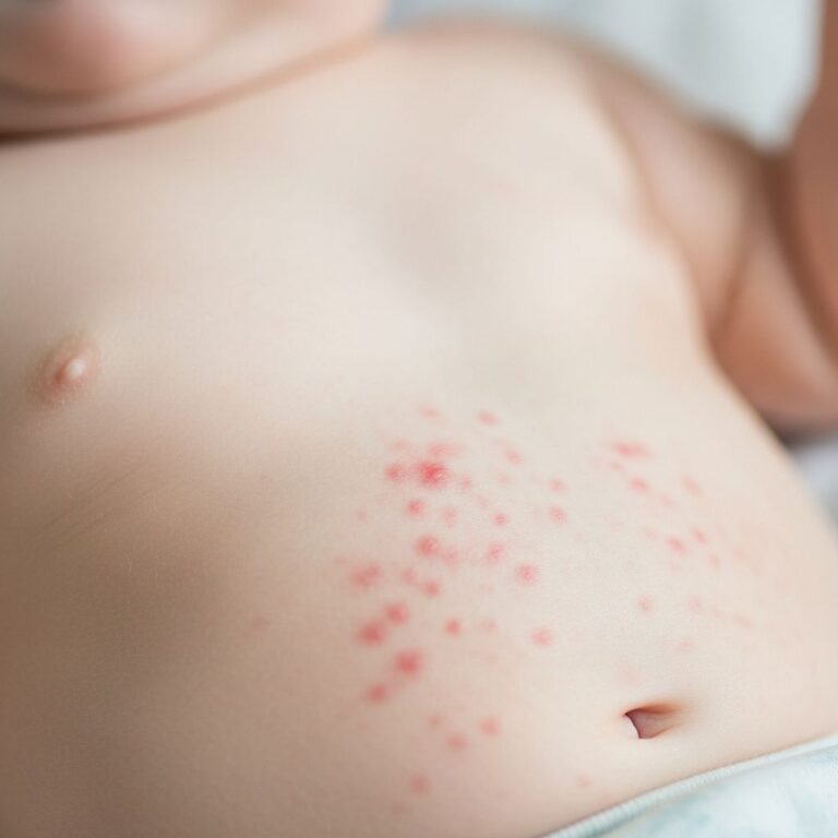
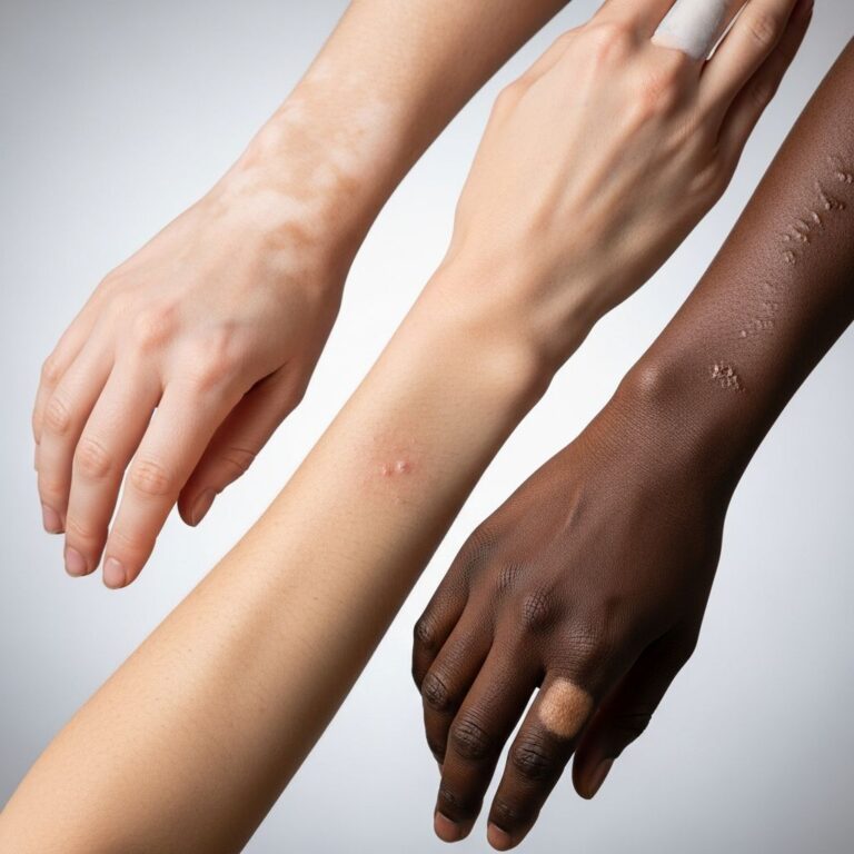
PCFCL classically presents with solitary or localized, slowly growing, asymptomatic lesions on the head and neck (60–70% of cases), trunk (20–30%), or upper extremities. Lesions appear as firm, pink to violaceous papules, plaques, or nodules, ranging from 1–10 cm in diameter. They are typically non-pruritic, smooth-surfaced, and lack ulceration.
- Common sites: Scalp, forehead, cheeks, back, chest
- Morphology: Nodules (most common), plaques, tumours; single (70%) or grouped (30%)
- Symptoms: Painless, non-itchy; rare B-symptoms (fever, night sweats, weight loss)
- Progression: Slow enlargement if untreated; spontaneous regression in <10%
Leg involvement, seen in 10–20% of cases, portends a worse prognosis with higher relapse rates. Multifocal lesions occur in 20–30%, usually regionalized.
Diagnosis
PCFCL is suspected clinically but definitively diagnosed via skin biopsy. Incisional or excisional biopsy of the most prominent lesion is preferred for adequate tissue sampling.
Histology
Microscopic features include nodular or diffuse dermal/subcutaneous infiltrates of centrocytes (small cleaved cells) ± centroblasts, mimicking reactive follicles but with atypical features: absent mantle zones, lack of tingible body macrophages, irregular outline, and BCL6+ cells extending beyond dendritic networks. A Grenz zone spares the epidermis.
Immunohistochemistry
| Marker | Expression in PCFCL |
|---|---|
| CD20, CD79a, PAX5 | Positive (B-cell) |
| CD10, BCL-6 | Positive (germinal centre) |
| BCL-2 | Usually negative |
| Ki-67 | Low (<30%) |
| MUM1, IgM | Negative |
Molecular studies
PCR for immunoglobulin gene rearrangements confirms clonality. Absence of t(14;18) IGH-BCL2 supports cutaneous origin. Next-generation sequencing may reveal characteristic mutations.
Staging investigations
- Complete physical exam including lymph nodes
- Blood tests: FBC, LDH, beta-2 microglobulin
- Imaging: PET-CT or CT if multifocal/large lesions
- Bone marrow biopsy (rarely indicated, <5% positive)
Differential diagnosis
Clinical mimics include basal cell carcinoma, amelanotic melanoma, or pseudolymphoma. Histological differentials require immunohistochemistry:
| Condition | Key Distinguishing Features |
|---|---|
| Follicular lymphoma (systemic) | t(14;18)+, BCL2+, extracutaneous disease |
| Primary cutaneous marginal zone lymphoma | Small lymphocytes/plasma cells, light chain restricted |
| Reactive lymphoid hyperplasia | Polyclonal, tingible body macrophages, polarized follicles |
| PCDLBCL, leg type | MUM1+, BCL2+, high Ki-67, leg location |
Management
Treatment is guided by lesion number, size, location, and patient factors. Observation may be appropriate for small, stable lesions. Complete remission is achieved in 90–100% with localized therapy.
Localized disease (single/grouped lesions)
- Radiotherapy: First-line; 24–30 Gy electrons, cure rates >95%
- Surgical excision: For small (<2 cm) lesions
- Intralesional steroids/rituximab: Alternatives
Multifocal disease
- Rituximab: IV or intralesional; response 90–100%
- R-CHOP: Rarely for aggressive/recurrent cases
- Other: Radiotherapy fields, IFN-alpha
Prognosis and follow-up
PCFCL has an excellent prognosis: 5-year survival >95% overall, 40–60% with leg involvement. Relapses occur in 30–50% (usually local skin), but rarely progress to systemic disease (<5%).
- Follow-up: Every 3–6 months initially; full skin/lymph node exam
- Monitoring: Clinical; PET-CT if relapse suspected
Frequently asked questions (FAQs)
Q: Is primary cutaneous follicle centre lymphoma curable?
A: Yes, localized PCFCL is often cured with radiotherapy or excision, achieving >95% remission. Multifocal disease relapses but remains indolent.
Q: What is the most common site for PCFCL?
A: Head and neck region, particularly scalp and forehead (60–70% of cases).
Q: Does PCFCL spread to internal organs?
A: Extremely rare (<5%); it is defined by lack of extracutaneous disease at diagnosis.
Q: Can PCFCL regress spontaneously?
A: Yes, in rare cases (<10%), but most lesions progress slowly if untreated.
References
- Primary Cutaneous Follicle Center Lymphoma — MD Searchlight. 2023. https://mdsearchlight.com/cancer/primary-cutaneous-follicle-center-lymphoma/
- Primary cutaneous follicle centre lymphoma — DermNet NZ. 2024-01-15. https://dermnetnz.org/topics/primary-cutaneous-follicle-centre-lymphoma
- Primary cutaneous follicle centre lymphoma (PCFCL) — SEER Cancer.gov. 2023-05-01. https://seer.cancer.gov/seertools/hemelymph/51f6cf5ae3e27c3994bd549b/
- Unusual Clinical Presentation of a Primary Cutaneous Follicle Center Lymphoma — PMC/NCBI. 2019-11-12. https://pmc.ncbi.nlm.nih.gov/articles/PMC6883459/
- Skin (cutaneous) B-cell lymphoma — Lymphoma Action. 2024. https://lymphoma-action.org.uk/information-and-support/types-lymphoma/skin-lymphoma/skin-cutaneous-b-cell-lymphoma
- Primary Cutaneous Follicle Center Lymphoma — StatPearls/NCBI Bookshelf. 2023-07-17. https://www.ncbi.nlm.nih.gov/books/NBK567743/
Similar Articles


Read full bio of Sneha Tete