Primary Dermal Melanoma: Definition, Diagnosis & Treatment
Rare dermal melanoma subtype with favourable prognosis compared to metastases, guiding targeted excision and surveillance.

Primary dermal melanoma (PDM) represents a rare and distinctive subtype of cutaneous melanoma characterized by its confinement exclusively to the dermis, without epidermal involvement. This condition poses unique diagnostic challenges as it mimics metastatic melanoma but demonstrates markedly superior prognosis, influencing treatment decisions toward curative intent rather than palliative approaches.
What is primary dermal melanoma?
Primary dermal melanoma arises as a primary lesion entirely within the dermal layer of the skin, lacking any junctional or in-situ component typically seen in conventional cutaneous melanomas. Histopathologically, it presents as a nodular proliferation of atypical melanocytes confined to the dermis, often resembling a nodular melanoma without surface involvement. This subtype has been recognized due to its unexpectedly favourable clinical behaviour, with survival rates significantly outperforming those of dermal metastases from cutaneous primaries.
The key distinction lies in its primary origin: PDM develops de novo in the dermis, potentially from immature melanocytes or regressed superficial components, rather than spreading from an epidermal primary. Studies from large melanoma databases, such as the Melanoma Research Database at Melanoma Institute Australia, confirm that PDM patients exhibit 5-year overall survival rates of 87.1% and melanoma-specific survival superior to both early-stage cutaneous melanomas and stage IV M1a disease. This evidence supports treating PDM as a true primary lesion amenable to aggressive local therapy.
Who gets primary dermal melanoma?
Primary dermal melanoma affects both sexes equally, with no gender predilection reported in the literature. The average age at diagnosis is approximately 51.2 years, though cases have been documented across a broad range from 8 to 83 years, indicating it can occur in paediatric and elderly populations alike. Ethnicity data remains limited, but no specific racial predispositions have been identified, unlike conventional melanomas which show higher incidence in fair-skinned individuals.
Risk factors mirror those of cutaneous melanoma, including UV exposure history, though the dermal confinement suggests possible alternative pathogenesis such as dermal melanocyte migration anomalies or prior trauma inducing regression of any epidermal component. Comprehensive clinicopathological review is essential, as demographic profiles do not strongly differentiate PDM from other melanomas.
What does primary dermal melanoma look like?
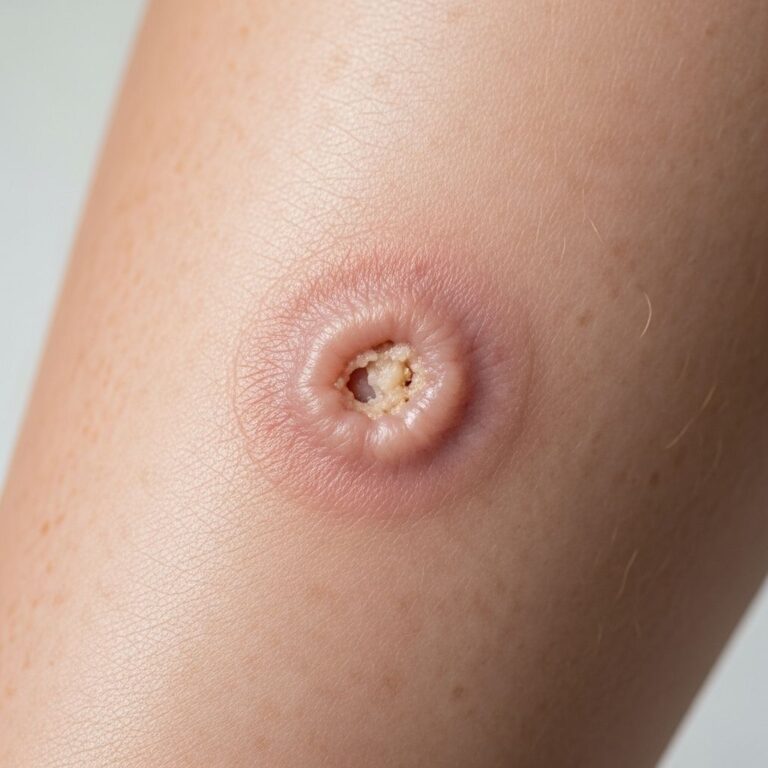
Clinically, primary dermal melanoma manifests as a solitary, often rapidly growing dermal nodule, typically asymptomatic but potentially showing changes in size, colour, or texture consistent with ABCDE criteria (Asymmetry, Border irregularity, Colour variation, Diameter >6mm, Evolving). The lesion appears as a firm, dome-shaped papule or nodule, ranging from 0.5 to several centimetres in diameter, with colours spanning pink, red, blue, or black due to dermal pigmentation.
Surface features may include ulceration or crusting from minor trauma, and regression phenomena like hypopigmentation can occur, mimicking healed lesions. Unlike superficial spreading melanoma, there is no visible epidermal plaque; the lesion feels deeply seated upon palpation. Dermoscopy may reveal atypical bluish structures or structureless areas without typical melanocytic networks, aiding in suspicion but not definitive diagnosis.
How is primary dermal melanoma diagnosed?
Diagnosis hinges on histopathological confirmation following excisional biopsy. Pathologists report a nodular melanoma confined to the dermis, lacking junctional activity or in-situ spread. Features include sheets or nests of epithelioid or spindle melanocytes with cytological atypia, mitotic activity, and possible necrosis. Immunohistochemistry shows positivity for S100, HMB45, Melan-A, and SOX10, confirming melanocytic origin.
Critical to diagnosis is ruling out metastasis via thorough clinical history (no prior primary), full skin examination, and imaging (CT/PET) to exclude occult primaries or distant disease. Sentinel lymph node biopsy (SLNB) may be performed if Breslow thickness exceeds 1mm or ulceration is present, mirroring cutaneous melanoma protocols. Clinicopathological correlation is paramount, as pure dermal location alone does not confirm primacy.
Differential diagnosis of primary dermal melanoma
The primary differential is cutaneous metastasis from an occult or regressed cutaneous melanoma, which shares identical histology but portends dismal prognosis (5-year survival ~52%). Other considerations include:
- Spitz naevus or atypical Spitz tumour: More common in young patients; lacks deep mitoses and shows maturation.
- Blue naevus or cellular blue naevus: Benign, symmetrical, with dendritic melanocytes and no atypia.
- Dermal naevocellular naevus: Lacks cytological atypia and mitoses; shows nevus nesting.
- Metastatic carcinoma or sarcoma: Distinguished by immunohistochemistry.
- Melanophagic dermatitis: Inflammatory response without neoplastic melanocytes.
Advanced molecular profiling, such as BRAF/NRAS/KIT mutations, may aid but is not routine. Prognostically, correct identification as PDM shifts management from systemic therapy to local excision.
Prognosis and survival of primary dermal melanoma
PDM confers an excellent prognosis, with literature reporting 3–5 year survival rates of 73–100%, far exceeding the 52% 5-year survival for cutaneous melanoma metastases. A landmark study of 62 PDM cases showed 5-year overall survival of 87.1% and superior outcomes versus stage I-II cutaneous melanomas (p=0.0002 overall survival) and stage IV M1a (p<0.0001). Median follow-up was 6.3 years, with disease-free survival comparable to early-stage primaries but better than metastatic controls.
Adverse factors include tumour thickness (>2mm), ulceration, high mitotic rate, and lymph node involvement, akin to conventional melanoma. Nonetheless, even thicker PDMs respond well to excision, with low metastatic potential. Compared to nodular melanoma (poorer prognosis due to vertical growth), PDM’s dermal restriction paradoxically protects.
| Stage/Comparison | 5-Year Survival | 10-Year Survival | Source |
|---|---|---|---|
| PDM | 87.1% | 74.2% (overall) | |
| Stage I-II Cutaneous | ~90-98% | 80-95% | |
| Stage IV M1a | <50% | N/A | |
| Cutaneous Metastasis | 52% | N/A |
Treatment and management of primary dermal melanoma
Management mirrors early-stage cutaneous melanoma: wide local excision (WLE) with 1-2cm margins based on tumour thickness, followed by reconstruction if needed. Sentinel lymph node biopsy is recommended for intermediate-thickness lesions (>1mm) to stage accurately. Unlike metastases, systemic therapy (immunotherapy or targeted agents) is reserved for documented nodal or distant relapse.
Follow-up entails regular dermatological surveillance, skin exams every 3-6 months initially, with imaging as indicated. Patient education on self-examination and sun protection is crucial. Recent advances in adjuvant checkpoint inhibitors for stage II melanoma may apply to high-risk PDM, though data is emerging. Surgery remains primary, with 5-year survival post-WLE approaching 95-100% for thin lesions.
Primary dermal melanoma progressing to metastatic disease
Though rare, PDM can metastasise, particularly if thick or ulcerated. Progression rates are low (<15% in cohorts), but nodal or distant spread warrants staging and multidisciplinary review. Upon metastasis, prognosis aligns more with stage III/IV disease, though initial dermal confinement may confer relative chemosensitivity. Vigilant follow-up mitigates this risk.
Frequently asked questions (FAQs) about primary dermal melanoma
Q: Is primary dermal melanoma a true primary cancer?
A: Yes, evidence shows PDM behaves as a primary lesion with superior survival to metastases, justifying curative excision.
Q: How does PDM differ from nodular melanoma?
A: Nodular melanoma involves epidermis with rapid vertical growth; PDM is purely dermal without junctional component.
Q: What is the survival rate for PDM?
A: 5-year survival is 87.1%, better than many stage I-II melanomas and vastly superior to metastases.
Q: Do I need lymph node biopsy for PDM?
A: Recommended if thickness >1mm or high-risk features present, per cutaneous melanoma guidelines.
Q: Can PDM be cured?
A: Most cases are curable with wide excision; metastasis is uncommon but possible in advanced lesions.
References
- Primary dermal melanoma: clinical behaviour, prognosis and implications for management – a retrospective study of 62 cases — Haydu et al. 2020-05-25. https://pubmed.ncbi.nlm.nih.gov/32417156/
- Primary dermal melanoma — DermNet NZ. 2023. https://dermnetnz.org/topics/primary-dermal-melanoma
- Prognosis of Patients With Primary Melanoma Stage I and II — Mohr et al. 2022. https://ascopubs.org/doi/10.1200/JCO.22.00202
- Prognosis and survival for melanoma skin cancer — Canadian Cancer Society. 2024. https://cancer.ca/en/cancer-information/cancer-types/melanoma-skin/prognosis-and-survival
- Melanoma Treatment – NCI — National Cancer Institute. 2025. https://www.cancer.gov/types/skin/patient/melanoma-treatment-pdq
Similar Articles


Read full bio of Sneha Tete