Protozoan Infections
Comprehensive guide to protozoan infections affecting the skin: causes, symptoms, diagnosis, and treatments.
Protozoan infections are caused by single-celled eukaryotic organisms known as protozoa, which can affect humans through various transmission routes including insect vectors, contaminated water, or direct contact. While many protozoan infections primarily target internal organs, some manifest prominently on the skin and mucous membranes, leading to distinctive dermatological conditions. These infections are more prevalent in tropical and subtropical regions but can occur worldwide due to travel and migration. Early recognition is crucial as skin lesions often provide the first clinical clue to systemic involvement.
What are they?
Protozoa are microscopic parasites that thrive in warm, moist environments and infect humans via fecal-oral route, arthropod bites, or sexual contact. Cutaneous involvement arises when protozoa invade skin tissues or migrate through them. Key examples include Leishmania species causing leishmaniasis, Trypanosoma species in trypanosomiasis, Entamoeba histolytica in amoebiasis, and Toxoplasma gondii in toxoplasmosis. These pathogens exploit host immune responses, leading to ulcers, nodules, or granulomatous lesions on the skin.
Who gets them?
Individuals in endemic areas, particularly those with poverty, poor sanitation, and proximity to vectors like sandflies or tsetse flies, are at highest risk. Immunocompromised patients, such as those with HIV/AIDS, are more susceptible to severe forms. Travelers to regions like South America, Africa, India, and the Middle East may acquire infections. Children and the elderly face higher complications due to weaker immunity.
Overview
- Leishmaniasis: Transmitted by sandfly bites; presents as cutaneous, mucocutaneous, or visceral forms with skin ulcers.
- Trypanosomiasis: African (sleeping sickness) via tsetse flies and American (Chagas disease) via triatomine bugs; causes chancre and Romana’s sign.
- Amoebiasis: Fecal-oral transmission; cutaneous form rare but destructive around perianal area.
- Toxoplasmosis: From cat feces or undercooked meat; skin involvement in immunocompromised.
These infections often require prolonged treatment and may be refractory.
Cutaneous leishmaniasis
Introduction
Cutaneous leishmaniasis (CL), also known as oriental sore or espundia, is caused by over 20 Leishmania species. It is the most common form, affecting millions annually in the Middle East, Central Asia, and Latin America.
Transmission
Phlebotomine sandflies transmit promastigotes during blood meals. Urbanization and deforestation increase vector exposure.
Clinical features
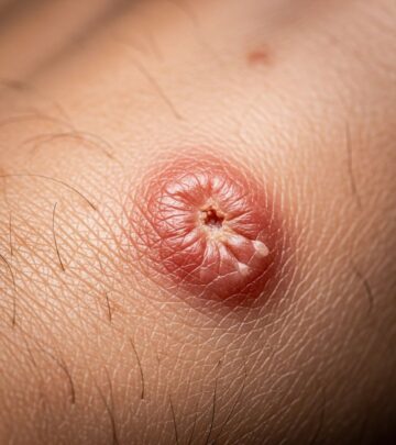
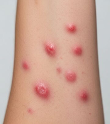
Lesions begin as pruritic papules at bite sites, evolving into nodules, plaques, or ulcers with raised borders and central crusting. Healing leaves atrophic scars. Satellite lesions or sporotrichoid spread may occur. Incubation: 2-24 weeks.
| Form | Features | Common Sites |
|---|---|---|
| Localized CL | Single ulcer | Exposed skin |
| Diffuse CL | Multiple nodules | Face, limbs |
| Mucocutaneous | Destructive erosions | Mouth, nose |
Diagnosis
Confirmed by skin biopsy showing amastigotes in macrophages (Giemsa stain), PCR, or culture on NNN medium. Montenegro skin test is supportive.
Management
Treatment depends on species and site. Options include intralesional antimonials, topical paromomycin, oral miltefosine, or systemic sodium stibogluconate (20 mg/kg/day IV/IM for 20-28 days). Amphotericin B for resistant cases. Cure rates high if treated early.
Leishmaniasis – American
Caused by L. braziliensis complex, this mucocutaneous form starts as cutaneous ulcers but progresses to nasopharyngeal destruction months to years later (espundia). Treatment mirrors CL but systemic therapy essential to prevent dissemination.
Visceral leishmaniasis
Primarily L. donovani or L. infantum; skin signs include hyperpigmentation or post-kala-azar dermal leishmaniasis (PKDL) with hypopigmented macules and nodules post-treatment.
Trypanosomiasis – African
Introduction
African trypanosomiasis (sleeping sickness) is caused by Trypanosoma brucei gambiense (West African, chronic) or T. b. rhodesiense (East African, acute). Endemic in sub-Saharan Africa.
Clinical features
Trypanosomal chancre: painful, indurated papule at bite site (1-2 weeks post-exposure). Followed by fever, lymphadenopathy, and Winterbottom’s sign (posterior cervical nodes). Late stage: meningoencephalitis.
Diagnosis and management
Diagnosis via blood smear, lymph node aspirate, or CSF for trypomastigotes. Treatment: suramin or pentamidine for early stage; melarsoprol or eflornithine for CNS involvement.
Trypanosomiasis – American (Chagas disease)
Caused by T. cruzi, transmitted by triatomine bugs. Acute: chagoma (unilateral periorbital edema, Romana’s sign) or erythema nodosum. Chronic: subcutaneous nodules (chagomas).
Diagnosis: serology, xenodiagnosis. Treatment: benznidazole or nifurtimox, most effective acutely.
Amoebiasis cutis
Rare extension of intestinal amoebiasis by E. histolytica. Perianal or genital ulcers: deep, ragged, base-covered with yellow slough, undermined edges. Painful, progressive.
Diagnosis: trophozoites in biopsy or exudate. Treatment: metronidazole 750 mg TDS for 10 days + diloxanide furoate.
Toxoplasmosis
T. gondii rarely causes primary skin lesions but in immunocompromised: roseola-like eruption, erythroderma, or urticaria. Congenital: blueberry muffin spots.
Diagnosis: serology (IgM/IgG). Treatment: pyrimethamine + sulfadiazine + folinic acid.
Differential diagnosis
| Condition | Key Differentiators |
|---|---|
| Pyoderma | Bacterial culture positive |
| Sporotrichosis | Lymphocutaneous spread, fungal elements |
| Blastomycosis | Broad-based buds on biopsy |
| Syphilis | Dark-field microscopy |
Disease severity
Mild: self-limiting nodules. Severe: mutilating mucocutaneous disease or dissemination in HIV.
Prognosis
Excellent for localized CL with treatment. Poor for untreated mucocutaneous or visceral forms.
Prevention
- Insect repellents, bednets.
- Vaccine research ongoing.
- Safe water, hygiene for amoebiasis.
- Avoid undercooked meat for toxoplasmosis.
Frequently Asked Questions
What causes protozoan skin infections?
Primarily vector-borne transmission by sandflies, tsetse flies, or triatomine bugs, or fecal-oral route.
Are protozoan infections contagious?
No direct person-to-person; require vectors or contamination.
How are they diagnosed?
Biopsy, PCR, culture, serology.
What is the treatment duration?
Often 3-4 weeks; refractory cases longer.
Can they be fatal?
Yes, untreated visceral or CNS involvement.
References
- Treatment of protozoan infections — PubMed/NCBI. 2004-11-01. https://pubmed.ncbi.nlm.nih.gov/15571500/
- Protozoan Infections of the Skin and Eyes — eCampus Ontario Pressbooks. 2023. https://ecampusontario.pressbooks.pub/microbio/chapter/protozoan-and-helminthic-infections-of-the-skin-and-eyes/
- Parasitic infections of the skin — Tropical Medicine. 2018-05. https://www.tropmed.org/wp-content/uploads/2018/05/chapter10-2.pdf
- Leishmaniasis — UF Health. 2024. https://ufhealth.org/conditions-and-treatments/leishmaniasis
- Parasitic Infection — Cleveland Clinic. 2023. https://my.clevelandclinic.org/health/diseases/24885-parasitic-infection
- Protozoan infections — DermNet NZ. 2024. https://dermnetnz.org/topics/protozoan-infections
Similar Articles


Read full bio of Sneha Tete


