Pseudoxanthoma Elasticum: Causes, Symptoms & Treatment
Understanding PXE: A comprehensive guide to this rare connective tissue disorder affecting skin, eyes, and cardiovascular health.

Pseudoxanthoma Elasticum: A Comprehensive Overview
Pseudoxanthoma elasticum (PXE) is a rare hereditary disorder of connective tissue that fundamentally affects the structure and function of elastic fibers throughout the body. This multisystem orphan disease clinically impacts the skin, eyes, and cardiovascular system with considerable morbidity and potential for serious complications. Characterized by the progressive fragmentation and calcification of elastic fibers, PXE presents a diagnostic challenge to healthcare providers due to its late-onset nature and variable clinical presentations. Clinicians first recognized PXE more than a century ago, yet many patients experience a prolonged diagnostic odyssey that can last a decade or longer.
What Causes Pseudoxanthoma Elasticum?
PXE is caused by mutations in the ABCC6 gene, which encodes a protein responsible for regulating calcium and inorganic phosphate transport. These pathogenic variants result in abnormal mineralization and calcification of elastic fibers in various tissues throughout the body. The condition follows an autosomal recessive inheritance pattern, meaning an affected individual must inherit two mutated copies of the gene—one from each parent. Unlike conditions with similar cutaneous findings, such as generalized arterial calcification of infancy (GACI) caused by mutations in the ENPP1 gene, PXE specifically involves ABCC6 gene mutations. The precise mechanism by which these genetic mutations lead to elastic fiber degradation and calcification continues to be an area of ongoing research in medical genetics and dermatology.
Clinical Manifestations of PXE
Skin Manifestations
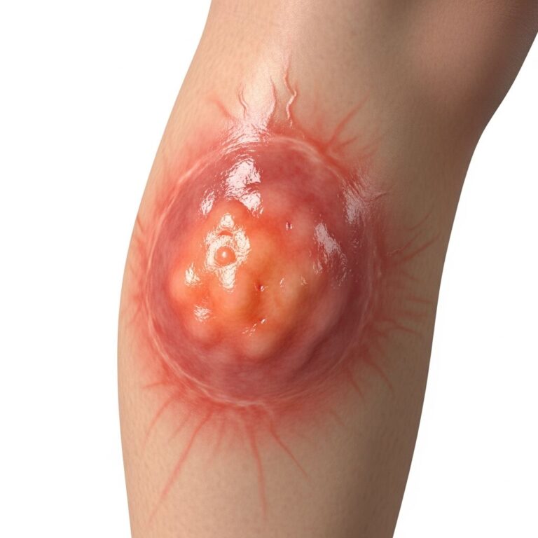
The primary cutaneous manifestation of PXE involves the appearance of small, discrete papules with a yellowish hue, often resembling xanthomas. These characteristic lesions typically develop initially on predilection sites, including the lateral neck, axillae, antecubital fossae, popliteal fossae, wrists, and groin. The lateral neck is frequently the first area of involvement, making it an important diagnostic site for clinical examination.
As the disease progresses, these individual papules slowly coalesce into larger plaques that eventually render the affected skin thick, grooved, stiff, and loose. The texture characteristically becomes similar to an orange peel or a plucked chicken, a feature described as peau d’orange. Although these skin findings are primarily of cosmetic concern, they represent an important early diagnostic indicator of the potential for more serious ocular and vascular complications. Notably, skin findings can be minimal or absent even in the presence of significant ocular or vascular manifestations, which underscores the importance of comprehensive screening in affected individuals.
Clinical manifestations can appear as early as the first or second decade of life, though early cutaneous findings are frequently subtle and often not recognized until the second or third decade. The change in appearance may be mild and overlooked during early childhood but becomes progressively more noticeable as the individual ages.
Ocular Manifestations
The eye involvement in PXE represents one of the most characteristic and clinically significant aspects of the disease. Angioid streaks (AS)—which are breaks in Bruch’s membrane due to calcification of the elastic lamina—are recognized as the most characteristic ocular finding in PXE. These streaks appear as linear breaks radiating from the optic disc and represent areas of weakness in the retinal structure.
More commonly observed than angioid streaks is a mottling peau d’orange retinal change, representing subconfluent calcification of Bruch’s membrane. Almost all patients will exhibit angioid streaks by the age of 30 years. While angioid streaks themselves are asymptomatic, choroidal neovascularization may develop within these fractures, leading to retinal hemorrhage and subsequent scarring with gradual vision loss.
Early ocular changes are typically visible only during ophthalmological examination, which may occur before noticeable skin changes develop. Later symptoms include distortion of vision and loss of central vision, which can significantly impact quality of life. For this reason, regular ophthalmological screening is essential for early detection and management of vision-threatening complications.
Cardiovascular Manifestations
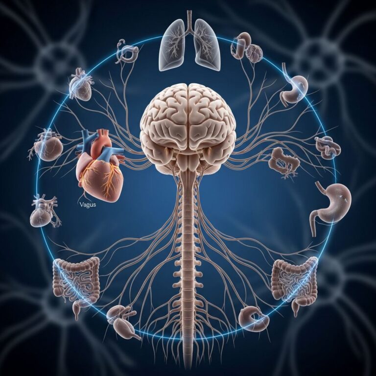
Cardiovascular manifestations of PXE result from calcification of the elastic media and intima of vessels, primarily affecting medium-sized vessels of the extremities, which leads to premature atherosclerosis. The clinical manifestations of vascular involvement are diverse and potentially serious:
- Intermittent claudication and angina pectoris
- Decrease or loss of peripheral pulses
- Renovascular hypertension
- Increased rates of mitral valve prolapse
- Myocardial infarction
- Stroke and transient ischemic attacks
- Cerebrovascular disease presenting as ministrokes and multi-infarct dementia
Coronary artery disease represents a significant complication, presenting with angina pectoris and less frequently myocardial infarction. Additionally, involvement of the vessels of the gastric and intestinal mucosa increases the incidence of gastrointestinal (GI) bleeding, including acute upper gastrointestinal hemorrhage and intestinal angina.
Gastrointestinal Complications
PXE can lead to serious gastrointestinal manifestations, primarily through calcification of blood vessels supplying the stomach and intestines. These vascular changes result in an increased incidence of gastrointestinal bleeding, which can be life-threatening and requires prompt medical intervention.
Diagnostic Criteria and Evaluation
Diagnostic Framework
The diagnosis of PXE requires a comprehensive approach combining clinical findings, histopathological examination, and genetic testing. A 2014 diagnostic criteria framework was established to standardize the diagnostic process and distinguish PXE from conditions with similar cutaneous findings.
Major Diagnostic Criteria
The 2014 diagnostic criteria include the following major criteria for definitive diagnosis:
- Cutaneous findings: Characteristic papules and plaques on flexural areas
- Histopathology: Elastic fiber fragmentation and calcification in the reticular dermis
- Ocular findings: Angioid streaks larger than one disc diameter in length or peau d’orange changes
- Genetic testing: Molecular genetic testing revealing the presence of biallelic ABCC6 pathogenic variants
Minor Diagnostic Criteria
The diagnostic framework also includes minor criteria that support the diagnosis:
- PXE in a first-degree relative
- Genetic test showing a pathologic variant in one allele of ABCC6
- PXE changes in the histopathology of nonlesional skin
Probable and Definitive Diagnosis
A probable diagnosis of PXE can be made with an affected first-degree relative plus either both major cutaneous criteria or one major ophthalmic criterion. Definitive diagnosis requires either two pathogenic mutations in the ABCC6 gene or ocular findings (angioid streaks greater than one disc diameter or peau d’orange in an individual under 20 years of age) together with characteristic skin findings and diagnostic histopathological changes.
Diagnostic Procedures
The diagnostic evaluation of PXE involves several complementary approaches:
- Clinical examination: Dermatological assessment of characteristic skin lesions, typically on the lateral neck and flexural areas
- Skin biopsy: Histopathological examination from affected lesions or from the lateral neck or flexural areas, stained with hematoxylin-eosin and special calcium stains to reveal calcium deposits, shortened and fragmented elastic fibers, and deformation of collagen fibers
- Ophthalmological examination: Fundoscopy to identify angioid streaks and peau d’orange changes; fluorescein fundus angiography or indocyanine green angiography may be performed for confirmation
- Genetic testing: Molecular testing to identify ABCC6 mutations
- Blood tests and imaging studies: CT of the heart and imaging of the brain to evaluate cardiovascular and cerebrovascular complications
Diagnostic Challenges and Timing
PXE presents significant diagnostic challenges, particularly because clinical manifestations are late-onset and early cutaneous findings are often subtle and frequently not recognized until the second or third decade of life. If the diagnosis of PXE is not definitive in suspected individuals under 30 years of age, the disease should be considered provisional, and clinical examinations should be repeated at 5-year intervals. This approach acknowledges the progressive nature of the disease and the gradual appearance of diagnostic features.
PXE-Like Syndromes
It is important to distinguish true PXE from PXE-like syndromes that may present with similar cutaneous changes but have different genetic causes. A PXE-like syndrome may be precipitated by long-term D-penicillamine use for the treatment of cystinuria or Wilson disease. Additionally, PXE-like cutaneous changes can be encountered in other ectopic mineralization disorders, including generalized arterial calcification of infancy (GACI) caused by mutations in the ENPP1 gene. Proper genetic testing and comprehensive evaluation are essential to distinguish these conditions and guide appropriate management strategies.
Management and Clinical Monitoring
While no cure currently exists for PXE, comprehensive management focuses on monitoring and treating the systemic complications that arise from elastic fiber calcification. Multidisciplinary care involving dermatologists, ophthalmologists, cardiologists, and gastroenterologists is essential to address the varied manifestations of the disease.
Regular ophthalmological screening is critical to detect early vision-threatening complications and implement preventive measures. Cardiovascular assessment should include evaluation for hypertension, coronary artery disease, and cerebrovascular disease. Gastrointestinal bleeding episodes require prompt medical intervention and may necessitate specialist referral. Genetic counseling is important for affected families, particularly given the autosomal recessive inheritance pattern and the implications for future generations.
Frequently Asked Questions
Q: At what age do PXE symptoms typically appear?
A: Clinical manifestations can appear as early as the first or second decade of life, though early cutaneous findings are often subtle and not recognized until the second or third decade. Ocular changes may develop by age 30, while skin findings become increasingly noticeable with age.
Q: How is PXE inherited?
A: PXE follows an autosomal recessive inheritance pattern, meaning an affected individual must inherit two mutated copies of the ABCC6 gene—one from each parent who may be carriers without symptoms.
Q: Can PXE be cured?
A: Currently, no cure exists for PXE. Management focuses on monitoring and treating complications through multidisciplinary care involving various specialists.
Q: What is the most characteristic ocular finding in PXE?
A: Angioid streaks, which are breaks in Bruch’s membrane due to calcification of the elastic lamina, are the most characteristic ocular finding. Almost all patients develop these by age 30.
Q: How often should individuals with PXE have ophthalmological screening?
A: Regular ophthalmological screening is essential, particularly in younger individuals, to detect early vision-threatening complications like choroidal neovascularization and retinal hemorrhage.
Q: What cardiovascular complications should PXE patients monitor?
A: PXE patients should be monitored for atherosclerosis, hypertension, coronary artery disease, stroke, myocardial infarction, and peripheral vascular disease due to elastic fiber calcification in blood vessels.
References
- Pseudoxanthoma elasticum — VisualDx. Accessed January 2026. https://www.visualdx.com/
- Pseudoxanthoma Elasticum: Diagnostic Features and Management — PubMed Central (PMC). 2014. https://pmc.ncbi.nlm.nih.gov/articles/PMC4219573/
- Pseudoxanthoma Elasticum – Symptoms, Causes, Treatment — National Organization for Rare Disorders (NORD). https://rarediseases.org/rare-diseases/pseudoxanthoma-elasticum-pxe/
- Pseudoxanthoma Elasticum — Merck Manuals. https://www.merckmanuals.com/home/children-s-health-issues/connective-tissue-disorders-in-children/pseudoxanthoma-elasticum
- Pseudoxanthoma Elasticum (PXE) — DermNet New Zealand. https://dermnetnz.org/topics/pseudoxanthoma-elasticum
- Pseudoxanthoma elasticum — MedlinePlus Genetics. U.S. National Library of Medicine. https://medlineplus.gov/genetics/condition/pseudoxanthoma-elasticum/
- Pseudoxanthoma elasticum — Cleveland Clinic Journal of Medicine, 2024. https://www.ccjm.org/content/93/1/13
Similar Articles


Read full bio of Sneha Tete