Pulmonary Embolism: Overview, Symptoms, and Treatment
Understanding pulmonary embolism: causes, symptoms, diagnosis, and evidence-based treatment options.

What Is Pulmonary Embolism?
A pulmonary embolism (PE) is a blood clot that travels through the veins and lodges in the pulmonary arteries of the lung, blocking blood flow. This life-threatening condition occurs when a clot—typically originating from a deep vein thrombosis (DVT) in the legs or arms—dislodges and travels through the circulatory system to the lungs. PE restricts blood flow, reduces oxygen levels in the lungs, and increases blood pressure in the pulmonary arteries, creating a medical emergency requiring prompt diagnosis and treatment.
Together, PE and DVT form the spectrum of venous thromboembolism (VTE), a significant cause of morbidity and mortality worldwide. PE is one of the most common heart and blood vessel diseases globally, ranking third behind heart attack and stroke. In the United States alone, approximately 900,000 people per year develop a PE, with an incidence of 60 to 120 cases per 100,000 people annually.
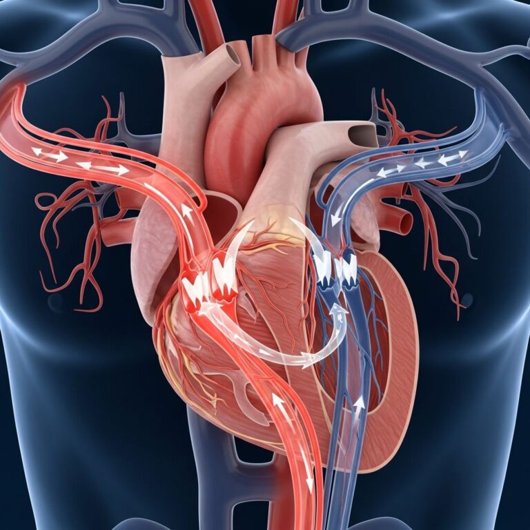
How Pulmonary Embolism Develops
Understanding the pathophysiology of PE helps explain why this condition is so serious. When a blood clot obstructs the pulmonary arteries, it impairs gas exchange in the lungs. The blockage creates a mismatch between ventilation and perfusion—alveolar ventilation continues normally, but pulmonary capillary blood flow decreases, leading to dead space ventilation and hypoxemia (low blood oxygen).
The body responds by releasing mediators such as serotonin, which cause vasospasm and further decrease pulmonary blood flow in unaffected areas. These inflammatory mediators also alter lung surfactant and stimulate the respiratory drive, resulting in hypocapnia and respiratory alkalosis.
Hemodynamically, PE increases pulmonary vascular resistance through two mechanisms: mechanical obstruction of the vascular bed by the thrombus and hypoxic vasoconstriction. When thromboembolic occlusion exceeds 30% to 50% of the total cross-sectional area of the pulmonary arterial bed, pulmonary artery pressure rises. This increased resistance raises right ventricular afterload, impairing right ventricular outflow and causing right ventricular dilation. Consequently, left ventricular filling decreases, reducing cardiac output and causing systemic hypotension and hemodynamic instability. Right ventricular failure due to acute pressure overload is the primary cause of death in severe PE.
Risk Factors for Pulmonary Embolism
Multiple factors increase the risk of developing a PE. These include:
- Genetic predispositions such as thrombophilia (a tendency for blood to clot abnormally)
- Prolonged immobility (long flights, bed rest, extended sitting)
- Recent surgery or trauma
- Malignancy (cancer)
- Pregnancy and the postpartum period
- Oral contraceptive use or hormone replacement therapy
- Heart or lung disease
- Obesity
- Smoking
- History of previous blood clots
If you have any of these risk factors and have experienced a blood clot, consult with your healthcare provider about preventive measures to reduce your PE risk.
Symptoms of Pulmonary Embolism
PE symptoms vary depending on the size and location of the clot. The most common symptoms include:
- Dyspnea (shortness of breath) — often acute and severe in central PE, but mild and transient in small peripheral PE
- Chest pain — particularly pleuritic chest pain (worsening with breathing or exertion)
- Cough
- Hemoptysis (coughing up blood)
- Presyncope or syncope (fainting or near-fainting)
- Tachycardia (rapid heart rate)
- Cyanosis (bluish skin discoloration) in massive PE
Notably, PE is a well-recognized cause of sudden cardiac arrest, accounting for approximately 8% of cases. In patients with preexisting heart failure or pulmonary disease, worsening dyspnea may be the only symptom. Some individuals with PE may experience no symptoms at all, making diagnosis more challenging.
Severe cases with massive PE may present with signs of acute right ventricular failure, including jugular venous distension, parasternal lift, a third heart sound, cyanosis, and shock. A concerning clinical sign is when a PE patient with initial tachycardia suddenly develops bradycardia or new broad complex tachycardia with right bundle branch block—this indicates right ventricular strain and impending shock.
Diagnosis of Pulmonary Embolism
Diagnosing PE requires a combination of clinical assessment and diagnostic testing. The diagnostic approach depends on the patient’s hemodynamic stability:
For Hemodynamically Stable Patients
The standard three-step evaluation includes:
- Clinical Probability Assessment: Healthcare providers use validated scoring systems such as the Wells criteria and Geneva criteria to estimate the likelihood of PE based on clinical presentation and risk factors.
- D-dimer Testing: A blood test measuring fibrin degradation products; negative results help exclude PE in low-probability patients.
- Chest Imaging: CT pulmonary angiography (CTPA) is the gold standard imaging modality, providing high sensitivity and specificity for PE detection.
For Hemodynamically Unstable Patients
Unstable patients with systolic blood pressure below 90 mm Hg may require bedside imaging for rapid diagnosis without waiting for formal testing.
Additional Diagnostic Tools
- Venous ultrasound to detect DVT in the lower extremities
- Electrocardiography (ECG) to assess for signs of right ventricular strain
- Chest X-ray to rule out alternative diagnoses
- Arterial blood gas analysis to evaluate oxygenation status
Treatment of Pulmonary Embolism
PE management combines supportive measures with anticoagulation therapy and, in severe cases, reperfusion strategies.
Anticoagulation Therapy
Anticoagulation is the mainstay of PE treatment, preventing clot enlargement and reducing recurrence risk. First-line therapy consists of direct oral anticoagulants (DOACs), including apixaban, edoxaban, rivaroxaban, and dabigatran, due to their efficacy and convenience. Traditional options include low-molecular-weight heparin (LMWH) and unfractionated heparin (UFH).
Reperfusion Strategies for Severe PE
For hemodynamically unstable patients (systolic blood pressure < 90 mm Hg), systemic thrombolysis is the recommended first-line therapy. Thrombolysis is associated with an absolute reduction in mortality of 1.6% (from 3.9% to 2.3%). Other reperfusion options include:
- Catheter-directed thrombolysis: Delivery of thrombolytic agents directly to the clot via catheter
- Catheter-directed therapy: Mechanical or pharmacomechanical approaches to remove or fragment the clot
- Surgical embolectomy: Surgical removal of the clot, reserved for specific clinical scenarios
Supportive Care
Patients receive oxygen therapy to maintain adequate oxygenation, hemodynamic support for blood pressure maintenance, and monitoring in an intensive care setting when appropriate.
Long-Term Management and Prevention of Recurrence
Long-term anticoagulation is essential to prevent PE recurrence, with duration tailored to individual risk factors and the provenance of the initial event (provoked versus unprovoked). Patients should maintain regular follow-up appointments with their healthcare provider to monitor treatment effectiveness and assess for complications.
Chronic thromboembolic pulmonary hypertension (CTEPH) represents a chronic manifestation of recurrent PEs and requires specialized management.
Prognosis and Outcomes
With timely diagnosis and appropriate treatment, PE is seldom fatal. Without treatment, however, PE is a very serious condition that can lead to permanent illness or death, with some patients dying suddenly within hours of PE onset.
Mortality rates vary by patient population. With current treatment approaches, PE is fatal in only 1% to 3% of patients in the United States, though mortality risk is higher in patients with preexisting heart or lung conditions. In untreated cases or those with delayed diagnosis, mortality rates are substantially higher.
Annual mortality from PE in the United States is estimated at 60,000 to 100,000 deaths per year. The prognosis depends on multiple factors, including the size of the blood clot, extent of blockage, overall health status, and cardiac function. Recovery time varies considerably; it can take months or years for a PE to resolve completely.
Comparison of PE Severity and Management Approaches
| PE Classification | Hemodynamic Status | Primary Treatment | Mortality Risk |
|---|---|---|---|
| Massive PE | Systolic BP < 90 mm Hg | Systemic thrombolysis | High (3.9% without treatment) |
| Submassive PE | Systolic BP ≥ 90 mm Hg with RV dysfunction | Anticoagulation ± catheter therapy | Moderate |
| Non-massive PE | Systolic BP ≥ 90 mm Hg without RV dysfunction | Anticoagulation (DOACs) | Low (< 1%) |
Frequently Asked Questions About Pulmonary Embolism
Q: What is the difference between DVT and PE?
A: DVT (deep vein thrombosis) is a blood clot in a deep vein, usually in the leg. PE occurs when a clot from a DVT breaks off and travels to the lungs. Both conditions are part of the same disease spectrum called venous thromboembolism (VTE).
Q: Can PE be prevented?
A: Yes, prevention involves avoiding prolonged immobility, staying active, maintaining a healthy weight, avoiding smoking, using compression stockings when recommended, and taking prescribed anticoagulants if you have risk factors or a history of blood clots.
Q: How long do you need to take anticoagulants after PE?
A: The duration depends on whether your PE was provoked (caused by a specific trigger like surgery) or unprovoked. Provoked PEs typically require 3 months of anticoagulation, while unprovoked cases may require longer durations. Your healthcare provider will determine the appropriate duration for your specific situation.
Q: Is PE always fatal if untreated?
A: Not all untreated PEs are immediately fatal, but the risk of death is significantly higher without treatment. Approximately 8% of sudden cardiac arrest cases are caused by PE. With treatment, survival rates exceed 97%.
Q: What should I do if I suspect I have a PE?
A: Seek immediate medical attention. Call emergency services or go to the nearest emergency department if you experience sudden shortness of breath, severe chest pain, fainting, or coughing up blood. PE is a medical emergency, and prompt treatment greatly reduces the chance of death.
Q: Can you fully recover from a PE?
A: Most people recover well with appropriate treatment. However, recovery time varies—complete resolution can take months or years. Some patients may experience long-term complications such as chronic thromboembolic pulmonary hypertension (CTEPH).
When to Seek Emergency Care
Seek immediate medical attention if you experience any of the following:
- Sudden onset of severe shortness of breath
- Severe chest pain, especially with breathing or exertion
- Fainting or severe lightheadedness
- Coughing up blood
- Rapid or irregular heartbeat
- Bluish skin discoloration
PE is a medical emergency. With timely diagnosis and treatment, survival is excellent. Delaying care increases the risk of serious complications and death.
References
- Acute Pulmonary Embolism — National Center for Biotechnology Information (NCBI) StatPearls. 2024. https://www.ncbi.nlm.nih.gov/books/NBK560551/
- Pulmonary Embolism: Symptoms, Causes & Treatment — Cleveland Clinic. 2024. https://my.clevelandclinic.org/health/diseases/17400-pulmonary-embolism
- Acute Pulmonary Embolism: A Review — JAMA (Journal of the American Medical Association). 2023. https://jamanetwork.com/journals/jama/article-abstract/2796942
- Disparities in Current Pulmonary Embolism Management and Outcomes — American Heart Association Circulation Journal. 2024. https://www.ahajournals.org/doi/10.1161/CIR.0000000000001306
Similar Articles


Read full bio of medha deb