Recessive X-Linked Ichthyosis: Symptoms, Diagnosis, Treatment
Genetic skin disorder causing dry, scaly skin in males due to steroid sulfatase deficiency and X-linked inheritance.

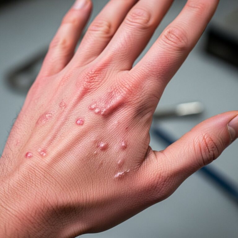
Recessive X-linked ichthyosis is a genetic skin disorder characterised by very dry skin with generalised fine or rhomboid, adherent, dark brown or light grey scaling, most prominent on the back of the neck, upper trunk, and extensor surfaces of the limbs. It typically presents at birth or within the first six months of life and primarily affects males due to its X-linked recessive inheritance pattern.
Introduction
Recessive X-linked ichthyosis, often shortened to X-linked ichthyosis (XLI), is an inborn error of metabolism resulting from a deficiency in the enzyme
steroid sulfatase (STS)
. This condition leads to the accumulation of cholesterol sulfate in the stratum corneum, impairing desquamation and causing retention hyperkeratosis with characteristic scaling. The disorder is not life-threatening but can significantly impact quality of life due to cosmetic concerns and associated extracutaneous features.The skin appears dirty due to the adherent scales, particularly giving a ‘dirty neck’ appearance. Scaling spares skin folds, palms, soles, face, and central trunk in many cases, and symptoms often improve with age and in warmer climates. Prevalence is estimated at 1 in 2,000 to 6,000 male births worldwide, affecting all ethnic groups equally.
Causes
X-linked ichthyosis arises from mutations or deletions in the
STS gene
located on the distal short arm of the X chromosome (Xp22.31). This gene encodes the steroid sulfatase enzyme, crucial for desulfating cholesterol sulfate to free cholesterol in the epidermis. Deficiency results in elevated cholesterol sulfate levels, which inhibit epidermal serine proteases like kallikrein 7, reducing corneocyte shedding and leading to hyperkeratosis.Most cases (about 90%) involve complete deletion of the STS gene, detectable by fluorescence in situ hybridisation (FISH) or array comparative genomic hybridisation (aCGH). Point mutations account for the remainder. Rarely, larger contiguous gene deletions encompass neighbouring genes, causing syndromic forms with additional features such as Kallmann syndrome, X-linked chondrodysplasia punctata, short stature, intellectual disability, or central nervous system anomalies.
Maternal 35S-cholesterol sulfate levels are markedly elevated in affected pregnancies, sometimes used historically for prenatal diagnosis, though genetic testing is now preferred.
Demographics
As an X-linked recessive disorder, XLI predominantly affects males, who inherit the mutated X chromosome from their carrier mothers. Females are typically asymptomatic carriers, though rare manifesting heterozygotes occur due to skewed X-inactivation. The condition spans all races and ethnicities with no predilection.
Incidence is approximately 1:2000-1:6000 newborn males. Family history may reveal multiple affected males on the maternal side. Carrier mothers often show subtle signs like mild scaling or comma-shaped corneal opacities.
Clinical Features
Symptoms usually manifest at birth or within the first weeks to months, sometimes with delayed labour due to reduced cervical dilatation from cholesterol sulfate accumulation. Initial presentation includes non-erythematous, polygonal, loosely adherent scales that evolve into finer, darker, tightly adherent rhomboid or plate-like scales.
Key cutaneous features:
- Generalised scaling, most pronounced on abdomen, back, buttocks, legs, and ‘dirty neck’.
- Spares flexures (popliteal/antecubital fossae), palms, soles, face, and scalp.
- Symptoms worsen in winter, improve in summer and with age (often milder post-puberty).
Extracutaneous manifestations (10-20% of cases):
- Asymptomatic punctate corneal opacities (comma-shaped in carriers).
- Cryptorchidism (undescended testes, ~20%).
- Attention deficit hyperactivity disorder (ADHD) or other neurodevelopmental issues (~10%).
Severity varies; mild cases may be subclinical, while severe ones cause significant scaling and social stigma.
Diagnosis
Diagnosis combines clinical findings, family history, and laboratory confirmation.
Clinical diagnosis: Supported by X-linked pedigree, typical scaling pattern, and onset.
Laboratory tests:
- STS enzyme assay on leukocytes, fibroblasts, or placenta (gold standard).
- Plasma cholesterol sulfate elevation (>10-fold).
- Skin biopsy: compact orthokeratotic hyperkeratosis, reduced or absent desmosomes.
- Genetic testing: STS deletion (FISH/MLPA) or sequencing (~95% detection rate).
Differentiate from autosomal recessive congenital ichthyosis (e.g., TGM1 mutations, collodion baby) via genetic testing. Slit-lamp ophthalmology for corneal opacities and testicular exam recommended.
Treatment
No curative therapy exists; management is symptomatic, focusing on hydration, keratolysis, and scale reduction without irritation.
Daily regimen:
- Bathe daily with lukewarm water and syndet bars or bath oils.
- Pat dry; apply emollients immediately (petrolatum, lanolin, or cholesterol-containing).
- Humectants/keratolytics: urea (10-20%), lactic acid (5-12%), salicylic acid (2-3%), glycolic acid.
Start low and slow to avoid stinging, especially in children. Combinations like urea+lactic acid enhance efficacy.
Advanced options:
- Topical retinoids (tazarotene 0.05% gel).
- Oral retinoids (acitretin) for severe refractory cases, intermittently in winter.
Address extracutaneous issues: urology for cryptorchidism, ophthalmology for opacities (rarely symptomatic), behavioural support for ADHD. Genetic counselling for families.
Outcome
Prognosis is excellent; no increased mortality or malignancy risk. Scaling persists lifelong but often attenuates with age. Psychological impact from appearance can be significant; support groups aid coping. Ongoing research explores gene therapy.
Treatment adherence improves cosmetics and quality of life.
Frequently Asked Questions (FAQs)
What causes recessive X-linked ichthyosis?
It is caused by mutations or deletions in the STS gene on the X chromosome, leading to steroid sulfatase deficiency and cholesterol sulfate accumulation.
Who does X-linked ichthyosis affect?
Primarily males; females are carriers. Incidence is 1:2000-6000 male births.
What does X-linked ichthyosis look like?
Dark brown/grey adherent scales on neck (‘dirty neck’), trunk, extensors; spares flexures, palms/soles.
How is it diagnosed?
Clinical features + STS enzyme assay, genetic testing for STS deletion.
Is there a cure?
No cure, but emollients, keratolytics (urea, alpha-hydroxy acids), and retinoids manage symptoms effectively.
Does it affect life expectancy?
No; it’s cosmetic with good prognosis, though extracutaneous issues like cryptorchidism need monitoring.
References
- Recessive X-linked ichthyosis — Orphanet. 2023. https://www.orpha.net/en/disease/detail/461
- X-Linked Ichthyosis — StatPearls, NCBI Bookshelf. 2023-10-01. https://www.ncbi.nlm.nih.gov/books/NBK448149/
- X-Linked Ichthyosis — First Skin Foundation. 2024. https://www.firstskinfoundation.org/types-of-ichthyosis/x-linked-ichthyosis
- Recessive X-linked ichthyosis — DermNet NZ. 2023. https://dermnetnz.org/topics/recessive-x-linked-ichthyosis
- Ichthyosis, X Linked — NORD (National Organization for Rare Disorders). 2023. https://rarediseases.org/rare-diseases/ichthyosis-x-linked/
Similar Articles


Read full bio of medha deb