Red Spot On Eye White: 6 Causes And What To Do
Discover causes, symptoms, and care for harmless red patches on the eye's white area that alarm many but often heal naturally.

A sudden red patch on the white part of your eye can be startling, but it’s often a benign condition known as subconjunctival hemorrhage where a tiny blood vessel bursts under the clear surface membrane. This issue affects people of all ages and typically resolves without intervention, though recognizing when it’s serious is key to eye protection.
Understanding the Basics of Eye Surface Bleeding
The eye’s outer layer includes the sclera, the firm white structure, covered by the conjunctiva, a thin, transparent tissue that keeps it moist and protected. When a small vessel beneath the conjunctiva ruptures, blood spreads out in that space, creating a vivid red area since the conjunctiva lets the color show through clearly. Unlike deeper eye bleeding, this doesn’t impact vision and feels mostly normal.
This phenomenon mimics a bruise on the skin: blood pools and gets reabsorbed over time by the body. The trapped blood can’t be washed away because it’s sandwiched between tissue layers, leading to a dramatic look that fades gradually.
Primary Triggers Behind Burst Eye Vessels
Several everyday factors can pressure these delicate vessels enough to cause rupture. Identifying patterns helps in avoidance.
- Sudden physical exertion: Intense coughing fits, explosive sneezes, forceful vomiting, or straining during heavy lifts or constipation spike blood pressure momentarily, popping vessels.
- Direct eye injury: Aggressive rubbing, especially with itchy or fatigued eyes, or impacts from sports, accidents, or debris can damage surface capillaries.
- Health-related pressures: Uncontrolled hypertension weakens vessel walls over time, making them prone to breaks.
- Blood-thinning influences: Aspirin, anticoagulants like warfarin, or supplements such as ginkgo increase bleeding risk by impairing clotting.
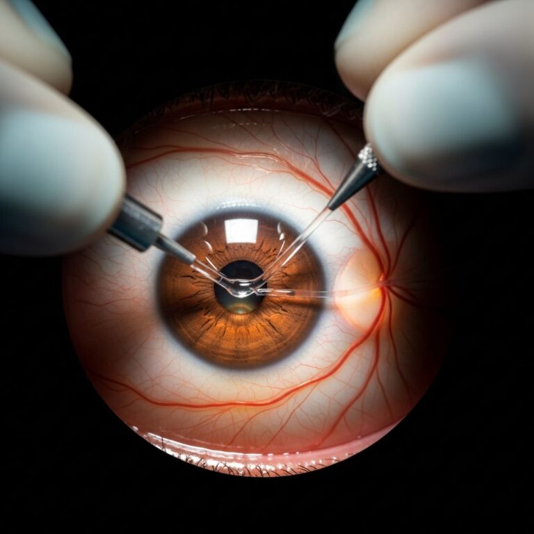
- Procedural aftereffects: Post-eye surgeries like cataract removal or laser treatments, plus irritation from poorly fitted contacts, contribute occasionally.
- Spontaneous occurrences: In seniors, aging makes vessels brittle, leading to breaks without obvious triggers; allergies, colds, or even alcohol can play roles too.
Not all cases tie to a clear event—up to half may be idiopathic, per clinical observations.
Recognizing Signs and What to Expect Visually
The hallmark is an abrupt bright red blotch on the sclera, often spotted in the mirror upon waking. It might start pinpoint-small or cover much of the white area.
| Symptom | Description | Common? |
|---|---|---|
| Red patch | Sudden vivid spot or spread on white eye area | Always |
| Pain | Absent in typical cases | Rare |
| Vision impact | No blur, loss, or field changes | None |
| Discomfort | Mild scratchiness or fullness, like minor irritation | Sometimes |
| Other effects | No tears, discharge, or light sensitivity | None |
Color evolution marks healing: red shifts to orange-yellow over 1-3 weeks as hemoglobin breaks down, fully clearing in 10-14 days usually. No itch or swelling accompanies pure cases.
Healing Timeline and Everyday Management
Patience is primary—most heal independently without meds or drops. Apply cool compresses briefly to ease any minor unease, avoid rubbing, and use artificial tears for dryness if present.
Steer clear of aspirin or thinners unless prescribed, and monitor blood pressure if recurrent. Contacts should pause on the affected eye until cleared. Full resolution averages 7-14 days, worsening slightly first as blood disperses.
When Professional Evaluation Becomes Essential
While alarming visually, isolated events rarely signal danger. Prompt checks are warranted if:
- Pain, swelling, or vision shifts like blur or floaters occur.
- Light sensitivity, discharge, or headaches join.
- Recurrences happen frequently, hinting at hypertension, diabetes, or clotting issues.
- Recent trauma or surgery precedes it, to exclude internal harm.
- Bleeding disorders or anticoagulant use complicates.
Deeper bleeds like retinal ones demand urgency due to vision threats from conditions such as vein occlusions or diabetic changes.
Preventive Strategies for Stronger Eye Vessels
Minimize risks through lifestyle tweaks:
- Control blood pressure with diet, exercise, and meds as advised.
- Gently manage allergies or colds to curb sneeze/cough force.
- Wear protective eyewear in risky activities.
- Ensure proper contact lens hygiene and fit.
- Limit rubbing; address dry eyes proactively.
- Consult on blood thinners for eye bleed history.
Regular eye exams catch vascular fragilities early, especially for those over 50 or with chronic conditions.
Distinguishing Surface from Serious Internal Bleeds
Surface subconjunctival types stay superficial and vision-neutral, unlike:
| Type | Location | Symptoms | Urgency |
|---|---|---|---|
| Subconjunctival | Under conjunctiva on sclera | Red patch only, no pain/vision loss | Low unless recurrent |
| Retinal | Within retina | Floaters, blur, vision loss | High |
| Hyphema | Front chamber | Blood visible in colored part, pain | High |
Quick differentiation by pros via exam prevents complications.
Frequently Asked Questions
Is a red spot on my eye white dangerous?
Typically no—it’s a harmless vessel burst that self-resolves, but watch for added symptoms signaling more.
How long until the red eye patch disappears?
1-2 weeks commonly, with color fading progressively.
Can I speed up healing of eye surface blood?
No treatments needed usually; compresses and tears help comfort, but time absorbs it.
Does high blood pressure cause frequent eye red spots?
Yes, it raises rupture risk; check levels if repeats occur.
Should I skip contacts with a burst eye vessel?
Yes, until healed to avoid irritation.
Is eye rubbing a common trigger for this?
Absolutely, especially vigorous sessions on irritated eyes.
Long-Term Eye Wellness After an Episode
Post-incident, prioritize hydration, UV protection, and balanced nutrition rich in vitamins A, C, E for vessel resilience. Annual optometrist visits track changes, particularly with family history of vascular issues. While single events dismiss easily, patterns prompt holistic health reviews—blood work, pressure logs—to safeguard vision longevity.
Empowerment comes from knowledge: that striking red doesn’t equate threat, yet vigilance ensures rare escalations get addressed swiftly. Maintain calm, observe progression, and engage care when deviations arise.
References
- Bleeding On The White Of The Eye: Causes, Symptoms & Treatment — AccuVision. Accessed 2026. https://www.accuvision.co.uk/news-and-blog/bleeding-on-the-white-of-the-eye/
- Understanding Eye Bleeding: Causes, Symptoms, and Treatments — NW Eye Clinic. Accessed 2026. https://nweyeclinic.com/understanding-eye-bleeding-causes-symptoms-and-treatments/
- Subconjunctival Hemorrhage: Care Instructions — MyHealth Alberta (Government of Alberta). Accessed 2026. https://myhealth.alberta.ca/Health/aftercareinformation/pages/conditions.aspx?hwid=uf7760
- Subconjunctival hemorrhage—potential causes and treatments — Acuvue. Accessed 2026. https://www.acuvue.com/en-us/eye-health/subconjunctival-hemorrhage/
- Subconjunctival Hemorrhage — Optometrists.org. Accessed 2026. https://www.optometrists.org/general-practice-optometry/guide-to-eye-health/eyes-and-allergies/subconjunctival-hemorrhage/
- Subconjunctival Hemorrhage — Willis Knighton Eye Institute. Accessed 2026. https://www.wkeyeinstitute.com/services-and-treatments/eye-diseases-and-conditions/subconjunctival-hemorrhage
- Subconjunctival Hemorrhage: Symptoms, Causes & Treatment — Cleveland Clinic. 2023-10-19. https://my.clevelandclinic.org/health/diseases/17713-subconjunctival-hemorrhage
Similar Articles


Read full bio of Sneha Tete