Renal Angiogram: Purpose, Procedure, and Results
Complete guide to renal angiogram: understand the procedure, preparation, and diagnostic benefits.

What Is a Renal Angiogram?
A renal angiogram is a specialized imaging test that allows physicians to visualize and evaluate the arteries supplying blood to your kidneys. During this procedure, a radiologist injects contrast dye into the renal artery and uses real-time X-ray imaging to observe how the dye flows through your kidney’s blood vessels. This diagnostic tool provides detailed information about the structure and function of renal blood vessels, helping doctors identify various vascular abnormalities and kidney-related conditions.
The renal angiogram is considered a minimally invasive procedure, meaning it involves only a small incision rather than a large surgical opening. Healthcare providers can use this test to examine whether your blood vessels are functioning properly and to detect any structural problems that might affect kidney health or overall cardiovascular function.
Why Might You Need a Renal Angiogram?
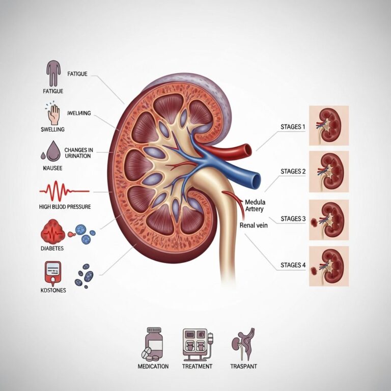
Your healthcare provider may recommend a renal angiogram to investigate various kidney and vascular conditions. The test helps doctors identify specific problems affecting blood flow to your kidneys and can guide treatment decisions. Common reasons for undergoing this procedure include:
- Suspected narrowing of a renal artery (renal artery stenosis) causing high blood pressure
- Bulging of a blood vessel, known as an aneurysm
- Blood vessel spasm (vasospasm)
- Blood clots blocking arteries that supply the kidney
- Abnormal connections between veins and arteries (fistulas)
- Suspected kidney tumors or cancers
- Unexplained kidney bleeding or hemorrhage
- Complications following kidney transplantation
- Noncancerous kidney tumors requiring evaluation
If other imaging tests such as CT scans or MRI examinations have not provided clear views of your kidney blood vessels, your doctor may recommend a renal angiogram for more detailed visualization. This procedure can also serve a therapeutic purpose, as doctors can sometimes treat certain conditions during the same procedure, such as widening narrowed arteries using angioplasty.
How to Prepare for Your Renal Angiogram
Proper preparation ensures the procedure runs smoothly and safely. Your healthcare provider will give you specific instructions tailored to your medical situation, and it is essential to follow these guidelines carefully.
Before Your Appointment
Your healthcare provider will explain the entire procedure to you and answer any questions you may have. You will need to sign a consent form authorizing the medical team to perform the angiogram. Take time to read this form carefully and ask for clarification on anything unclear.
Inform your doctor about any medications you are taking, including prescription drugs, over-the-counter medications, supplements, and herbal products. Your doctor may ask you to discontinue certain medications, particularly blood thinners like aspirin or ibuprofen, several days before the procedure. Tell your healthcare provider if you are pregnant, may be pregnant, are breastfeeding, or have allergies to X-ray contrast dye or any medications.
You will likely need to fast before the procedure, meaning you should not eat or drink anything for a specified period. Your healthcare provider will inform you of the exact fasting duration, which could range from several hours to overnight. This precaution prevents complications if sedation is needed during the test.
Pre-Procedure Testing
Your healthcare provider may order blood tests before the procedure to assess your kidney function and determine how quickly your blood clots. These results help the medical team prepare for any potential complications and ensure you are a good candidate for the procedure. Additional blood work may be necessary depending on your medical history and current health status.
Day of the Procedure
On the day of your renal angiogram, wear comfortable, loose-fitting clothing that you can easily remove. You will change into a hospital gown and lie on an X-ray table. A nurse or technician will establish an intravenous (IV) line in your arm to administer fluids and medications that help you relax during the procedure. The medical team will connect you to an electrocardiogram (ECG) monitor to record your heart’s electrical activity throughout the test and monitor your heart rate, blood pressure, and breathing rate.
The radiologist will examine the pulses in the area below where the contrast dye will be injected, typically in your groin area. They will mark these pulse points so that staff can check your circulation after the procedure. This ensures that the injection site does not compromise blood flow to your limb.
What to Expect During the Procedure
Understanding what happens during your renal angiogram can help reduce anxiety and allow you to cooperate fully with the medical team.
Local Anesthesia and Access
The technician will clean and shave a small area of your groin where the catheter will be inserted. The radiologist will then inject local anesthetic medicine to numb this area completely. You will feel slight pressure as the physician places a needle into an artery in your groin, though you should not experience significant pain due to the numbing medication. In some cases, an artery in the elbow area may be used instead of the groin.
Catheter Placement
Once the area is numb, the radiologist passes a thin wire through the needle into the artery, then removes the needle and inserts a long, thin, flexible tube called a catheter through the same opening. Using fluoroscopy, a real-time X-ray imaging technique, the physician carefully guides the catheter into the aorta, the main blood vessel leaving the heart, and then into the renal arteries.
You may feel some pressure or mild discomfort as the catheter moves through your arteries, but you should not experience sharp pain. It is important to remain still during this phase and follow the radiologist’s instructions, which may include holding your breath for 10 to 25 seconds at a time to obtain clear images.
Contrast Dye Injection and Imaging
Once the catheter is properly positioned in the renal artery, the radiologist injects contrast dye through the catheter. This special dye makes your blood vessels visible on X-ray images, as normal blood vessels do not appear on regular X-rays. As the dye flows through your kidney’s blood vessels, the radiologist takes multiple sets of X-ray images to capture different phases of blood flow.
The first set of images shows the arteries themselves, allowing the doctor to see their structure and identify any narrowing, blockages, or aneurysms. The second set of images captures blood flow through the smaller blood vessels called capillaries and the veins. Depending on the specific condition being evaluated, the radiologist may inject contrast dye multiple times to obtain comprehensive imaging from different angles.
During the injection of contrast dye, you may experience a warm or flushing sensation. Some patients report a slight metallic taste in their mouth. These sensations are normal and temporary. You might also feel the urge to urinate as the dye passes through your kidneys, but this is not actual urination and does not require concern.
Completion and Hemostasis
Once the radiologist has obtained all necessary images, they carefully remove the catheter from your artery. To prevent bleeding from the insertion site, the physician applies firm, direct pressure to the area for approximately 15 minutes. Some facilities use mechanical compression devices to maintain consistent pressure.
After the bleeding has stopped, the medical team applies a sterile dressing to the injection site. The radiologist may place a heavy object or sandbag over the site for a period of time to help prevent bleeding and reduce the risk of blood collecting under the skin, a condition called a hematoma. You will then be moved to a recovery area where you can rest and be monitored.
After Your Renal Angiogram
Recovery after a renal angiogram is generally quick, though certain precautions are necessary during the immediate post-procedure period.
Recovery Period
In the recovery area, nurses will monitor your vital signs, including blood pressure, heart rate, and oxygen levels. You should plan to rest for several hours after the procedure. Many patients feel drowsy or fatigued after receiving sedative medications, so it is important to have someone available to drive you home, as you will not be permitted to drive yourself on the day of the procedure.
You must keep your leg or arm straight for 4 to 6 hours following the angiogram to prevent bleeding from the catheter insertion site. Avoid strenuous activity, heavy lifting, and intense exercise for at least a few days after the procedure. Your doctor will provide specific guidance about when you can resume normal activities.
Managing the Injection Site
The insertion site may feel tender or show minor bruising, which is normal. Keep the area clean and dry, and watch for signs of infection or excessive bleeding. You may shower or bathe 24 hours after the procedure, but avoid submerging the site in water for extended periods until it has fully healed. Do not remove the dressing until instructed to do so.
Medication and Follow-Up
Your doctor may recommend pain relief medication if you experience discomfort at the injection site. You should resume taking your regular medications as directed by your healthcare provider unless you were specifically instructed to discontinue them. Drink plenty of water and other fluids for the next 24 hours to help flush the contrast dye from your system and protect your kidney function.
Contact your healthcare provider immediately if you experience excessive bleeding, severe pain, signs of infection such as fever or increased warmth at the site, numbness or tingling in your leg, or any other concerning symptoms. Your doctor will schedule a follow-up appointment to discuss the results and recommend any necessary treatment.
Understanding Your Results
The radiologist will analyze the images obtained during your renal angiogram and prepare a report describing their findings. Your healthcare provider will review these results with you and explain what they mean for your health.
Normal Results
Normal results indicate that your renal arteries are appropriately sized, unobstructed, and show good blood flow to both kidneys. The blood vessels appear uniform without narrowing, bulging, or blockages. Normal results suggest that your kidneys have adequate blood supply and that vascular disease is not causing your symptoms.
Abnormal Results
Abnormal findings may include renal artery stenosis, an abnormal narrowing of the artery that can cause high blood pressure and reduced kidney function. Your doctor might identify an aneurysm, which is an abnormal bulging or weakening of the arterial wall. The test can also reveal blood clots blocking blood flow, fistulas representing abnormal connections between arteries and veins, or active bleeding within the kidney.
If your angiogram shows kidney tumors, your doctor will determine whether they are cancerous or benign and discuss treatment options. The findings will guide your healthcare team in developing an appropriate treatment plan, which may include medications, angioplasty to widen narrowed vessels, stent placement to maintain vessel opening, or other interventions.
Risks and Complications
While renal angiography is generally considered safe, all medical procedures carry some risk. Potential complications are usually rare and minor but can occasionally be serious. These may include allergic reactions to the contrast dye, kidney damage particularly in patients with pre-existing kidney disease, bleeding or hematoma at the injection site, infection, blood clots, artery damage, and stroke in rare cases. Your healthcare provider will discuss these risks with you before the procedure and explain how they will minimize potential complications based on your individual medical situation.
Frequently Asked Questions About Renal Angiograms
Q: How long does a renal angiogram procedure take?
A: The procedure typically takes 30 to 60 minutes from start to finish, though this can vary depending on the complexity of your case and whether therapeutic interventions are performed simultaneously.
Q: Is the renal angiogram procedure painful?
A: The procedure should not be painful due to local anesthesia numbing the insertion site. You may feel pressure, mild discomfort, or a sensation of movement, but pain indicates a problem and should be reported to the radiologist immediately.
Q: Can a renal angiogram be used to treat conditions in addition to diagnosing them?
A: Yes, renal angiography can be combined with therapeutic procedures such as angioplasty to widen narrowed arteries or stent placement to maintain vessel opening, treating the condition during the same procedure.
Q: Will I need to stay overnight in the hospital after a renal angiogram?
A: Most patients go home the same day after several hours of observation. Hospital admission is typically not necessary unless complications develop or additional treatments are performed.
Q: When will I receive my results?
A: The radiologist usually completes the initial analysis within 24 hours. Your healthcare provider will schedule an appointment to discuss the results and recommend next steps in your care.
References
- Renal Angiogram — University of Rochester Medical Center. Accessed 2025. https://www.urmc.rochester.edu/encyclopedia/content?contenttypeid=92&contentid=p07721
- What to Know About Renal Arteriography — WebMD. Accessed 2025. https://www.webmd.com/a-to-z-guides/what-to-know-renal-arteriography
- Renal Arteriography Information — Mount Sinai. Accessed 2025. https://www.mountsinai.org/health-library/tests/renal-arteriography
- Renal Angiogram for Kidney Cancer — Stanford HealthCare. Accessed 2025. https://stanfordhealthcare.org/medical-conditions/cancer/kidney-cancer/kidney-cancer-diagnosis/renal-angiography.html
Similar Articles


Read full bio of medha deb