Retinopathy: Causes, Symptoms, and Treatment Options
Understanding retinopathy: From risk factors to prevention and comprehensive treatment strategies.

Understanding Retinopathy
Retinopathy is a progressive eye disease that affects the retina, the light-sensitive tissue at the back of the eye responsible for vision. This condition occurs when blood vessels in the retina become damaged, leading to vision problems and potentially blindness if left untreated. Retinopathy is particularly common among individuals with diabetes and represents one of the leading causes of vision loss in working-age adults worldwide.
The retina serves as a critical window into overall systemic health, and understanding retinopathy is essential for maintaining long-term vision and general well-being. Recent research has demonstrated that retinal imaging can predict the future risk of developing not only eye diseases but also cardiac, pulmonary, metabolic, and neuropsychiatric conditions. This underscores the importance of regular eye examinations and early detection of retinal abnormalities.
What is Diabetic Retinopathy?
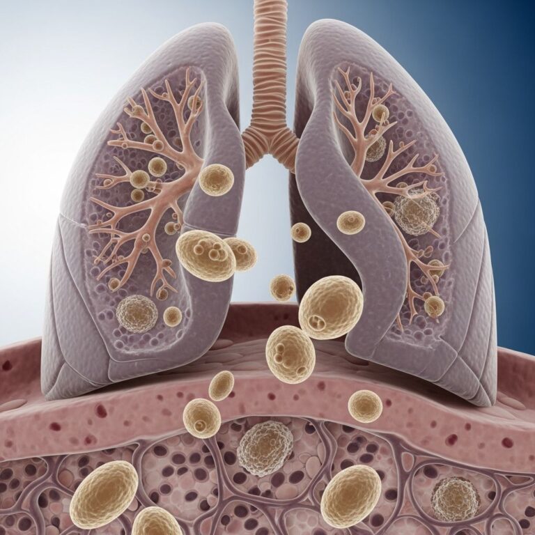
Diabetic retinopathy is the most common form of retinopathy and occurs as a complication of diabetes mellitus. This condition develops when high blood sugar levels damage the small blood vessels in the retina. The abnormal changes in these vessels lead to swelling, increased permeability, or abnormal growth of retinal blood vessels. As the disease progresses, these damaged vessels may leak fluid, causing macular edema, which affects central vision and can result in significant vision loss.
According to the World Health Organization, diabetic retinopathy causes blindness in almost 5 million people worldwide, making it a significant global health concern. In the United States, diabetic retinopathy is the leading cause of blindness in working-age adults, representing a critical public health challenge that demands attention to prevention and early intervention.
Risk Factors and Causes
Several factors increase the risk of developing retinopathy, with diabetes being the primary risk factor. The longer an individual has diabetes and the less controlled their blood sugar levels are, the higher their risk of developing retinopathy. Additional risk factors include:
- High blood pressure (hypertension)
- High cholesterol levels
- Pregnancy complications and gestational diabetes
- Smoking and tobacco use
- Poor kidney function
- Age and duration of diabetes
- Genetic predisposition and family history
Chronic hyperglycemia, or persistently elevated blood sugar, has devastating effects on blood vessels throughout the body, including those in the eyes. Individuals who depend on insulin injections to control their blood sugar are particularly susceptible to developing vascular problems in the eye. Research has identified 259 genetic loci associated with retinal thickness, suggesting that genetic factors play an important role in determining retinal health and susceptibility to retinal diseases.
Stages of Retinopathy
Diabetic retinopathy progresses through several stages, and early detection is crucial for preventing vision loss. Understanding these stages helps patients and healthcare providers implement appropriate interventions at each level of disease progression.
Nonproliferative Diabetic Retinopathy (NPDR)
This is the earliest stage of diabetic retinopathy. In this stage, small blood vessels in the retina become weakened and develop tiny outpouchings called microaneurysms. Fluid may leak from these damaged vessels, causing retinal swelling and the formation of hard exudates (yellowish deposits). Many people with NPDR experience no symptoms initially, though some may notice mild vision changes.
Proliferative Diabetic Retinopathy (PDR)
This advanced stage occurs when the retina becomes oxygen-deprived due to extensive blood vessel damage. The eye responds by growing new blood vessels, a process called neovascularization. However, these new vessels are abnormal and fragile, making them prone to bleeding. Bleeding into the vitreous (the gel-like substance filling the eye) can cause floaters, blurred vision, and potentially severe vision loss if bleeding is extensive.
Symptoms and Warning Signs
Early-stage retinopathy often produces no noticeable symptoms, which is why regular eye screenings are essential for individuals with diabetes. As the disease progresses, the following symptoms may develop:
- Floaters (small spots or lines floating in the visual field)
- Blurred or distorted central vision
- Dark or empty areas in the visual field
- Difficulty reading or performing detailed tasks
- Vision loss, particularly in peripheral areas
- Redness or irritation of the eye
- Eye pain (in advanced cases with bleeding)
Vision loss associated with retinopathy can be gradual or sudden, depending on the type and severity of retinal damage. Macular edema, which involves fluid accumulation in the macula (the central part of the retina responsible for detailed vision), is particularly likely to cause noticeable vision changes because it directly affects the area used for reading and recognizing faces.
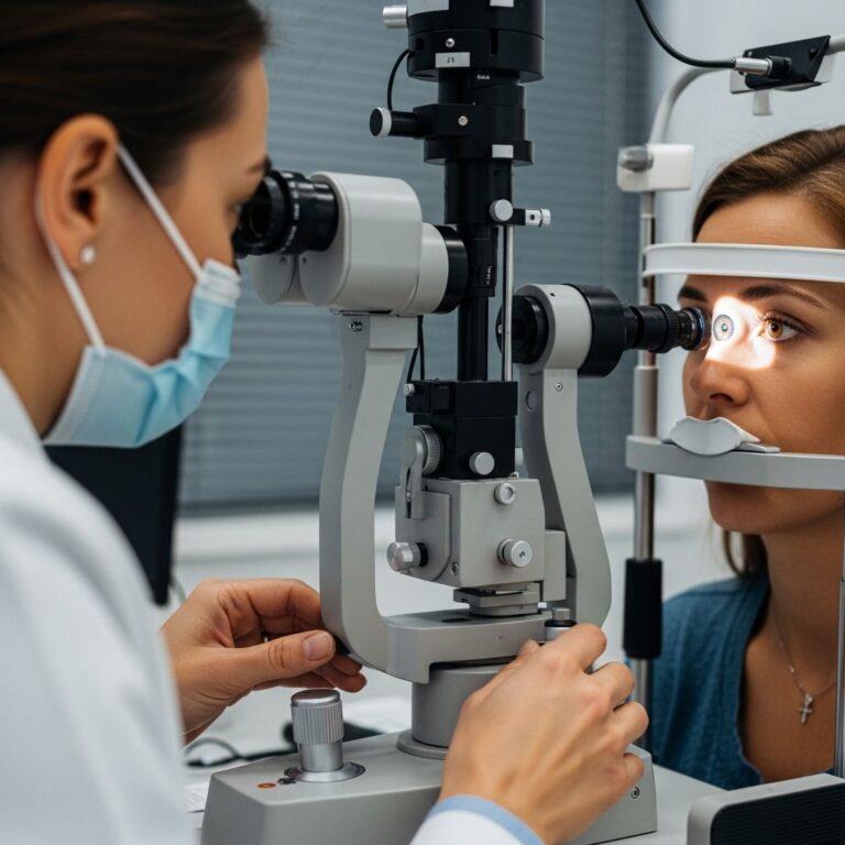
Diagnosis and Screening
Early detection of retinopathy significantly improves treatment outcomes and prevents vision loss. Ophthalmologists employ several diagnostic methods to identify and assess retinopathy:
Dilated Eye Examination
During a dilated eye exam, an ophthalmologist instills dilating drops to widen the pupil and examine the retina directly. This allows visualization of microaneurysms, hemorrhages, hard exudates, and other signs of retinal damage. The dilated examination remains the gold standard for detecting retinopathy and assessing its severity.
Optical Coherence Tomography (OCT)
OCT is a non-invasive imaging technique that provides detailed cross-sectional images of retinal layers. This technology is particularly useful for detecting macular edema and assessing the thickness of different retinal layers. Recent research has shown that OCT imaging can help predict future disease risk by identifying associations between retinal layer thickness and various ocular and systemic diseases. OCT has become a routine procedure in ophthalmology clinics and provides objective measurements of retinal health.
Fundus Photography
Color photographs of the retina create a permanent record of retinal appearance and changes over time. These images help ophthalmologists monitor disease progression and are valuable for comparing results of follow-up examinations.
Fluorescein Angiography
In this procedure, a fluorescent dye is injected into the bloodstream, and photographs are taken as the dye circulates through retinal blood vessels. This technique reveals areas of vascular leakage, ischemia (insufficient blood flow), and abnormal vessel growth.
Treatment Options
Treatment of retinopathy depends on the stage and severity of the disease. Advances in treatment over the last two decades have significantly improved outcomes for patients with diabetic retinopathy.
Blood Sugar Control
The foundation of retinopathy management is maintaining optimal glycemic control. Studies have demonstrated that intensive blood sugar management substantially reduces the risk of developing retinopathy and slows its progression in individuals who already have the condition. Working closely with an endocrinologist or diabetes specialist to achieve target blood sugar levels is critical.
Blood Pressure and Cholesterol Management
Controlling blood pressure and cholesterol levels further reduces the risk of retinopathy progression. Medications such as ACE inhibitors and statins have been shown to provide additional protective benefits beyond their primary effects on blood pressure and cholesterol.
Anti-VEGF Injections
Vascular endothelial growth factor (VEGF) inhibitors have become first-line therapy for diabetic macular edema and are also used for proliferative diabetic retinopathy. These medications are injected directly into the eye and work by reducing abnormal blood vessel growth and vascular permeability. Common anti-VEGF agents include bevacizumab, ranibizumab, aflibercept, and brolucizumab. These treatments have demonstrated superior efficacy compared to older laser-based approaches and are considered safe and effective options for preserving and restoring vision in diabetic patients.
Laser Photocoagulation
Laser treatment uses focused beams of light to seal leaking blood vessels and destroy abnormal new vessels. While less commonly used as first-line therapy than anti-VEGF injections, laser photocoagulation remains an important treatment option, particularly for certain presentations of proliferative diabetic retinopathy.
Vitrectomy
This surgical procedure involves removal of the vitreous gel from the eye when severe vitreous hemorrhage (bleeding) obscures vision or when proliferative tissue has become extensive. Vitrectomy allows restoration of clarity to the optical media and removal of scar tissue that may have developed.
Emerging Therapies
Research continues to identify novel therapeutic approaches for retinopathy. Recent discoveries, such as the identification of plasma kallikrein as a VEGF-independent mediator of diabetic macular edema, have led to the development of injectable and oral compounds currently in clinical trials. These developments promise additional treatment options for patients who may not respond adequately to existing therapies.
Prevention Strategies
Preventing retinopathy is far more effective than treating established disease. Key prevention strategies include:
- Maintaining tight glycemic control with a target HbA1c level determined by your physician
- Regular blood pressure monitoring and management to maintain optimal levels
- Annual comprehensive dilated eye examinations
- Maintaining a healthy diet rich in antioxidants and omega-3 fatty acids
- Regular physical activity and exercise
- Avoiding smoking and limiting alcohol consumption
- Managing kidney function and addressing kidney disease if present
- Maintaining healthy cholesterol levels through diet and medication as needed
The Role of Retinal Imaging in Disease Prediction
Emerging research demonstrates that retinal imaging extends beyond simply detecting retinopathy. Advanced analysis of retinal images can predict future risk of developing various ocular and systemic diseases, including glaucoma, macular degeneration, cardiac disease, pulmonary disease, metabolic disorders, and neuropsychiatric conditions. This capability suggests that routine retinal imaging might be leveraged more broadly for preventive medicine and early intervention across multiple medical specialties.
Artificial intelligence algorithms are being developed to analyze retinal images and predict disease progression and treatment response using multimodal retinal imaging. These advances may enable ophthalmologists to identify high-risk patients earlier and implement preventive strategies before vision loss occurs.
Impact on Quality of Life
Retinopathy significantly impacts the functional capacity and quality of life of affected individuals. Vision loss makes it difficult to perform daily activities, maintain employment, drive safely, and maintain independence. The psychological burden of progressive vision loss and the anxiety associated with potential blindness add to the overall impact of this disease.
Frequently Asked Questions
Q: Can retinopathy be cured?
A: While retinopathy cannot be completely cured, progression can be halted or slowed significantly through tight blood sugar control, blood pressure management, and appropriate medical or surgical interventions. Early detection and treatment are essential for preserving existing vision.
Q: How often should people with diabetes have eye exams?
A: The American Diabetes Association recommends that individuals with type 1 diabetes have an initial dilated eye examination shortly after diagnosis and at least annually thereafter. Those with type 2 diabetes should have an initial exam at the time of diagnosis and annually or more frequently if retinopathy is detected.
Q: Is retinopathy preventable?
A: Yes, retinopathy can be largely prevented through maintaining optimal blood sugar control, managing blood pressure and cholesterol, and receiving regular eye screenings. Studies show that intensive glycemic control reduces the risk of developing retinopathy by up to 76%.
Q: Does retinopathy always lead to blindness?
A: No. With early detection, appropriate treatment, and good disease management, most people with retinopathy maintain useful vision. However, if left untreated, advanced retinopathy can lead to severe vision loss or blindness.
Q: Can retinopathy develop without diabetes?
A: While diabetic retinopathy is the most common form, retinopathy can develop from other conditions including hypertension, kidney disease, blood disorders, and cancer treatment. However, diabetes remains the leading cause of retinopathy in developed countries.
Q: Are newer anti-diabetes medications beneficial for retinopathy?
A: Recent research is examining the effects of newer-generation anti-diabetes medications on retinopathy progression. Comprehensive blood sugar management with appropriate medications, combined with treatment of other risk factors, provides the best protection against retinopathy development and progression.
References
- Phenome- and genome-wide analyses of retinal optical coherence tomography images identify links between ocular and systemic health — Zekavat SM, et al., Mass Eye and Ear/Broad Institute. January 24, 2024. https://www.science.org/doi/10.1126/scitranslmed.adg4517
- Diabetic Eye Disease Center of Excellence — Department of Ophthalmology, Harvard Medical School. 2024. https://eye.hms.harvard.edu/diabetes
- The Functional Burden of Diabetic Retinopathy in the United States — PubMed Central, National Institutes of Health. 2021. https://pubmed.ncbi.nlm.nih.gov/33914161/
Similar Articles


Read full bio of medha deb