Retinopathy of Prematurity: Causes, Symptoms & Treatment
Understanding ROP: A guide to causes, detection, and treatment options for premature infants.
Retinopathy of Prematurity: Overview and Impact
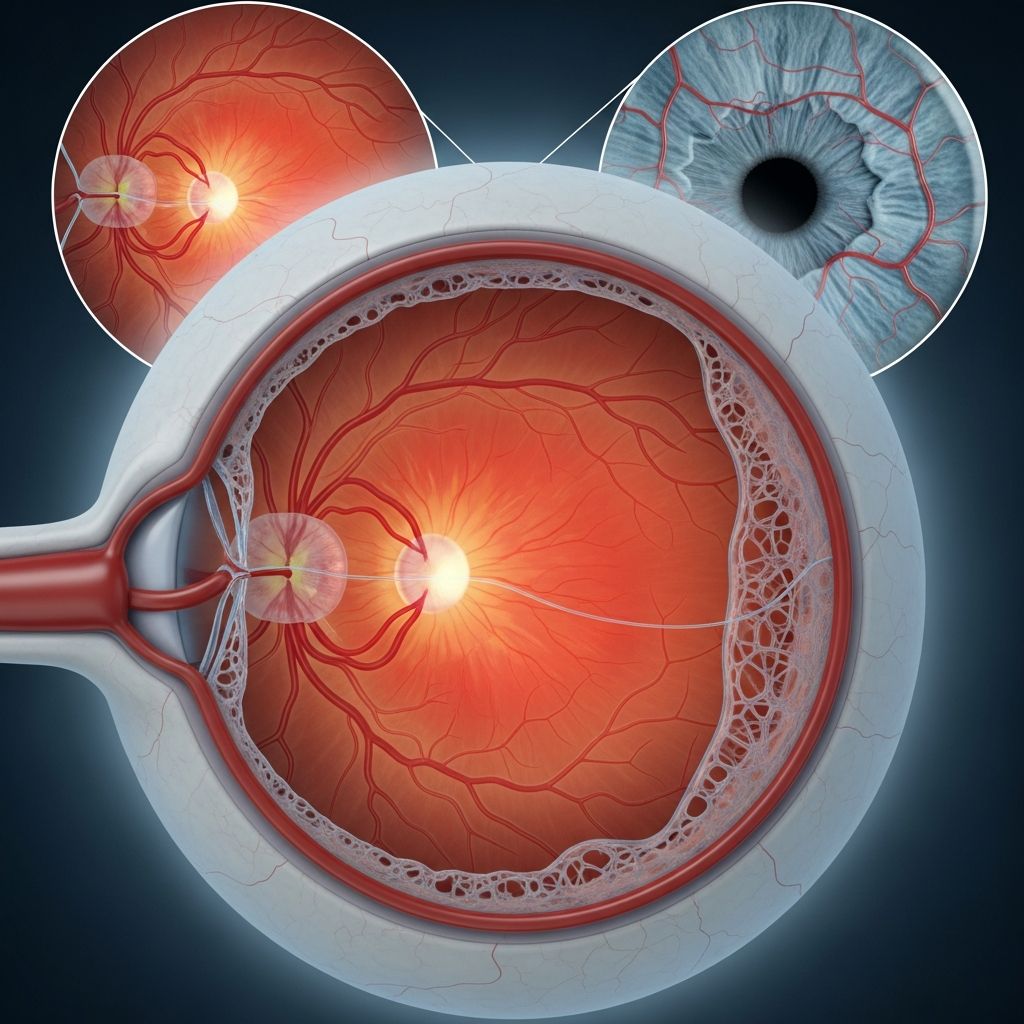
Retinopathy of prematurity (ROP) is a potentially blinding eye disease that develops in premature infants when abnormal blood vessels form in the retina, the light-sensitive tissue lining the back of the eye. This condition occurs when the normal development of retinal blood vessels is disrupted by early birth, leading to uncontrolled and abnormal vessel growth. In the most severe cases, ROP can result in bleeding, scarring, and retinal detachment—conditions that may cause irreversible vision loss or complete blindness. As a leading cause of preventable childhood blindness worldwide, ROP remains a significant concern in neonatal care, particularly as medical advances allow increasingly premature infants to survive.
Understanding the Cause and Development of ROP
During normal fetal development, the retina’s blood vessels begin to grow at approximately 16 weeks of gestation and continue developing until several weeks after birth. This carefully orchestrated process ensures proper oxygen and nutrient delivery to the developing retina. However, when an infant is born prematurely, this developmental process is interrupted, causing blood vessel growth to become irregular and abnormal. The factors that trigger this abnormal growth are multifaceted and involve both physiological stress and medical interventions necessary to keep premature infants alive.
Primary Risk Factors for ROP Development
The risk of developing ROP is directly correlated with the degree of prematurity and birth weight. Several established risk factors significantly increase the likelihood of ROP:
– Low birth weight below 1,500 grams (approximately 3.3 pounds), with smaller infants facing substantially higher risk- Birth at 32 weeks of gestation or earlier, with increased risk at earlier gestational ages- Prolonged exposure to high oxygen concentrations during neonatal intensive care, which can be highly toxic to developing blood vessels- Sepsis and other serious infections acquired during the neonatal period- Chronic lung disease requiring extended respiratory support- Inadequate nutrition and poor growth during critical developmental periods- Other systemic complications requiring intensive medical intervention
The combination of extreme prematurity and medical interventions, while necessary for survival, creates a challenging clinical situation where the benefits of life-sustaining treatment must be carefully balanced against the risk of developing ROP.
Recognizing Symptoms and Clinical Presentation
In its early stages, ROP typically produces no visible signs or symptoms. The only reliable method of detecting early-stage disease is through careful ophthalmologic examination. However, as the condition progresses to more severe stages, external symptoms may become apparent. These visible signs indicate advanced disease that warrants immediate attention:
– Leukocoria or white pupils, sometimes described as a white reflection in the pupil- Nystagmus, characterized by abnormal involuntary eye movements- Strabismus, commonly known as crossed eyes or eye misalignment- Severe myopia or nearsightedness, which may develop even in mild cases- In severe cases, eye redness, swelling, and discharge may occur
Parents and caregivers should be aware that the absence of visible symptoms does not indicate absence of disease. This is why routine screening by pediatric ophthalmologists is essential for all at-risk infants.
Classification: The Five Stages of ROP
Retinopathy of prematurity is classified into five distinct stages based on the severity and location of abnormal blood vessel growth and retinal changes. Understanding these stages helps guide treatment decisions and prognosis:
Stage 1: Mild Abnormality
Blood vessel growth is mildly abnormal, and the condition typically demonstrates a favorable natural history. Most infants with Stage 1 ROP recover completely without any medical intervention, maintaining normal vision throughout life. Regular monitoring remains important to ensure the condition does not progress.
Related Stories


Stage 2: Moderate Abnormality
Blood vessel growth becomes more noticeably abnormal, but the prognosis remains generally favorable. Similar to Stage 1, most babies with Stage 2 ROP will recover without requiring medical treatment. However, more frequent follow-up examinations are typically recommended to detect any progression.
Stage 3: Severe Abnormality
At this stage, blood vessel growth is significantly abnormal, and medical treatment often becomes necessary. Stage 3 represents a critical threshold where intervention can prevent progression to more severe, sight-threatening stages. Treatment decisions at this stage are crucial for preserving vision.
Stage 4: Partial Retinal Detachment
The developing scar tissue begins to contract, causing partial pulling away of the retina from the back wall of the eye. This partial detachment indicates progression to a more serious stage where the risk of vision loss increases substantially. Specialized retinal surgery may be considered at this stage.
Stage 5: Total Retinal Detachment
Complete separation of the retina from the eye wall occurs, resulting in irreversible vision loss or blindness. At this advanced stage, even aggressive surgical intervention may not restore vision. Prevention through early detection and treatment of earlier stages is paramount.
Screening and Early Detection
Early detection of ROP is the cornerstone of prevention of vision loss. Specialized screening programs ensure that at-risk infants receive timely examination before significant damage occurs. A pediatric ophthalmologist uses dilating eye drops to examine the interior of the baby’s eye, looking for characteristic changes in retinal blood vessels and tissue. Digital photography of the retina may be performed to document findings and track changes over time.
The initial screening typically occurs four to six weeks after birth in most at-risk infants. The specific timing may be adjusted based on individual risk factors. If findings are concerning, follow-up examinations are scheduled at intervals determined by the severity of findings, ranging from weekly to every other week. This close surveillance allows ophthalmologists to detect disease progression and intervene at the optimal time.
Treatment Options and Interventions
Treatment decisions for ROP depend on disease stage, severity of findings, and risk for progression. It is important to note that approximately 90% of infants with ROP improve spontaneously without treatment and achieve normal vision. However, the remaining 10% require intervention to prevent permanent vision loss.
Observation and Monitoring
For early-stage disease (Stages 1 and 2) with stable or regressing findings, careful observation with frequent ophthalmologic examinations is the standard approach. This conservative management strategy avoids unnecessary treatment while ensuring early detection of any progression that would warrant intervention.
Laser Therapy (Photocoagulation)
Laser therapy has been the gold standard treatment for advanced ROP for several decades. During this procedure, focused laser beams are carefully applied to specific areas of the retina, intentionally creating small, controlled scars to stop the growth of abnormal blood vessels. The laser destroys the peripheral areas of the retina where abnormal vessels are growing, effectively stopping progression of the disease. Laser therapy is typically performed in an operating room under appropriate anesthesia and sedation. While effective, laser therapy does result in permanent loss of peripheral vision, though central vision is usually preserved.
Intravitreal Injections
Newer treatment approaches involve injecting medication directly into the eye to slow or stop abnormal blood vessel growth. These medications inhibit vascular endothelial growth factor (VEGF), a key protein driving abnormal vessel formation. This treatment is gaining preference among many ophthalmologists due to several advantages: it is less invasive, can be performed at the bedside, is painless, and may allow more normal blood vessel development compared to laser therapy. Research continues to evaluate long-term outcomes and potential side effects of this approach. In some cases, injections are combined with laser therapy for optimal results.
Vitrectomy Surgery
For advanced disease with retinal detachment (Stages 4 and 5), vitrectomy may be recommended. This surgical procedure involves removing the gel-like substance filling the eye (vitreous humor) and carefully releasing traction on the detached retina. Vitrectomy is typically performed by retinal specialists and may be combined with other techniques such as lensectomy or silicone oil placement. While surgery may reattach the retina, visual function at this stage is often severely compromised.
Long-Term Follow-Up and Management
Children with a history of ROP require ongoing ophthalmologic follow-up extending well beyond the acute disease period. Even after ROP has resolved, these children have increased risk for refractive errors such as myopia and hyperopia, strabismus, and other structural eye abnormalities. Some infants develop chronic eye complications related to their ROP history. Regular comprehensive eye examinations throughout childhood and into adulthood help detect and manage these associated conditions early, optimizing visual development and educational outcomes.
Prevention Strategies
Reducing Premature Birth
The most effective strategy for preventing ROP is preventing premature birth itself. Obstetric interventions to prolong pregnancy when possible, and when delivery cannot be delayed, maternal administration of corticosteroids to accelerate fetal lung maturation may reduce both prematurity-related complications and the risk of severe ROP.
Careful Oxygen Management
Given that excessive oxygen exposure is a known risk factor for ROP, careful regulation of oxygen administration in neonatal intensive care is critical. Modern neonatal care employs pulse oximetry and blood gas monitoring to maintain oxygen saturation within target ranges that optimize survival while minimizing ROP risk. This balance requires vigilant attention from experienced neonatal teams.
Optimal Neonatal Care
Comprehensive neonatal intensive care addressing infection prevention, adequate nutrition, and management of respiratory and metabolic complications reduces overall morbidity and likely decreases ROP risk. Access to high-quality neonatal care has been shown to reduce both the incidence and severity of ROP in premature infant populations.
Frequently Asked Questions
Q: Will my premature baby definitely develop retinopathy of prematurity?
A: No. Not all premature infants develop ROP. The risk depends on the degree of prematurity, birth weight, and other medical factors. However, all infants at risk should receive screening examinations to detect the condition early if it does develop.
Q: What is the prognosis for a baby diagnosed with early-stage ROP?
A: Most infants with early-stage ROP (Stages 1 and 2) have an excellent prognosis, with spontaneous resolution and normal vision development in the majority of cases. Close monitoring ensures early detection of any progression requiring intervention.
Q: Are there any side effects from ROP treatment?
A: Laser therapy may result in some permanent peripheral vision loss, though central vision is typically preserved. Injection therapy is generally well-tolerated with minimal side effects, though long-term data continues to accumulate. Your ophthalmologist can discuss specific risks and benefits for your child’s situation.
Q: How long will my baby need follow-up eye exams?
A: Intensive screening continues until the retina has fully matured and ROP risk has passed, typically around 65 weeks postmenstrual age. Afterward, periodic comprehensive eye examinations throughout childhood are recommended to monitor for associated refractive errors and other complications.
Q: Can ROP be prevented?
A: While ROP cannot always be prevented, its incidence and severity can be reduced through careful oxygen management, infection prevention, optimal nutrition, and prenatal interventions to reduce premature birth when possible. Early detection and timely treatment of symptomatic disease prevent most cases of severe vision loss.
References
- Retinopathy of Prematurity: Causes, Symptoms and Treatment — Orbis International. Accessed December 2025. https://www.orbis.org/en/avoidable-blindness/retinopathy-of-prematurity
- Retinopathy of Prematurity: Causes & Treatment Options — Cleveland Clinic. Accessed December 2025. https://my.clevelandclinic.org/health/diseases/17430-retinopathy-of-prematurity
- Retinopathy of Prematurity (ROP) — Nemours KidsHealth. Accessed December 2025. https://kidshealth.org/en/parents/rop.html
- Retinopathy of Prematurity — Phoenix Children’s Hospital. Accessed December 2025. https://phoenixchildrens.org/specialties-conditions/retinopathy-prematurity
- Retinopathy of Prematurity: Treatment & FAQs — University of Virginia Children’s Hospital. Accessed December 2025. https://childrens.uvahealth.com/conditions/retinopathy-prematurity
- Retinopathy of Prematurity (ROP) — University of Michigan Health. Accessed December 2025. https://www.uofmhealth.org/our-care/specialties-services/retinopathy-prematurity-rop
- Retinopathy of Prematurity (ROP) — Boston Children’s Hospital. Accessed December 2025. https://www.childrenshospital.org/conditions/retinopathy-prematurity-rop
Similar Articles






Read full bio of medha deb