Rosai-Dorfman Disease Pathology: Diagnosis & Treatment Guide
Comprehensive pathology overview of Rosai-Dorfman disease, a rare histiocytic disorder with hallmark emperipolesis and varied clinical presentations.

Rosai-Dorfman disease (RDD), also known as sinus histiocytosis with massive lymphadenopathy, is a rare, benign histiocytic proliferative disorder primarily affecting lymph nodes but frequently involving extranodal sites such as the skin, soft tissues, and central nervous system.
Originally described in 1969, RDD predominantly impacts children and young adults with a male predominance, presenting as bilateral painless cervical lymphadenopathy often accompanied by fever, leukocytosis, and elevated ESR. Extranodal involvement occurs in up to 40-50% of cases, with cutaneous manifestations more common in older females.
Clinical Features
RDD classically manifests with massive, bilateral cervical lymphadenopathy in 80-90% of patients, though mediastinal, axillary, and inguinal nodes can also be affected. Constitutional symptoms like fever, night sweats, and weight loss are common in nodal disease. Extranodal RDD, seen in ~40% of cases, may occur with or without lymphadenopathy and involves diverse sites including skin (10%), CNS (<5%), orbits, salivary glands, bone, and lungs.
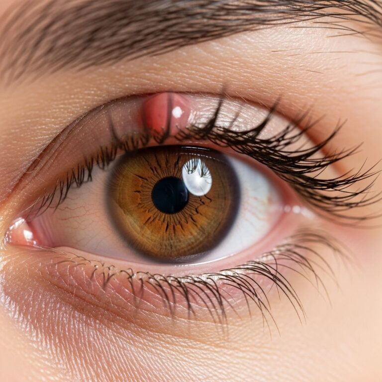
Cutaneous RDD typically presents in adults as solitary or multiple nodules, plaques, papules, or ulcerated lesions on the trunk, extremities, or face. Orbital involvement causes proptosis, eyelid swelling, or diplopia. CNS lesions mimic meningiomas with dural-based masses causing headaches or seizures.
- Nodal RDD: Painless, massive cervical nodes; chronic relapsing course.
- Extranodal RDD: Skin (papules/nodules), soft tissue masses, CNS dural lesions.
- Risk factors: Male predominance (1.5:1), peak age 5-15 years; familial forms linked to SLC29A3 mutations.
Histology
The histopathological hallmark of RDD is emperipolesis, where intact lymphocytes, plasma cells, neutrophils, and other inflammatory cells are phagocytosed but remain viable within the abundant cytoplasm of large histiocytes. These histiocytes are epithelioid or spindled with pale eosinophilic cytoplasm, vesicular nuclei, and prominent nucleoli, expanding lymph node sinuses or infiltrating extranodal tissues like dermis/subcutis with sclerosis.
In lymph nodes, histiocytes distend sinuses amid a mixed inflammatory background of lymphocytes, plasma cells (often IgG4-rich), neutrophils, and eosinophils. Fibrosis and vascular proliferation are prominent in regressed or chronic lesions. Cutaneous RDD shows dense dermal/subcutaneous infiltrates with sclerosis, multinucleated histiocytes, and abundant plasma cells, lacking the sinusoidal pattern of nodal disease.
Figure 1: Low-power view showing diffuse histiocytic infiltrate in dermis with sclerosis (H&E).
- Key histologic features:
- Sheets of large histiocytes with emperipolesis (hallmark).
- Pale/watery cytoplasm, hypochromatic nuclei.
- Polymorphous infiltrate: lymphocytes, plasma cells, neutrophils, eosinophils.
- Sclerosis and fibrosis, especially in skin/soft tissue.
Cytology
Fine-needle aspiration (FNA) or imprints reveal scattered large histiocytes with emperipolesis amid lymphocytes and plasma cells. Histiocytes show ample granular eosinophilic cytoplasm engulfing intact inflammatory cells. This finding, though characteristic, requires IHC confirmation as emperipolesis is not entirely specific.
Histochemistry
Limited role; periodic acid-Schiff (PAS) may highlight histiocyte cytoplasm, but immunohistochemistry is essential for diagnosis.
Immunohistochemistry
RDD histiocytes are strongly positive for S100, CD68, and CD163, with variable CD14 positivity. They are negative for CD1a, CD207 (langerin), and pan-T cell markers, distinguishing RDD from Langerhans cell histiocytosis (LCH). Cyclin D1 and IgG4 may be positive in plasma cells, hinting at IgG4-related disease overlap.
| Marker | RDD Histiocytes | LCH | Langerhans Cells |
|---|---|---|---|
| S100 | Strong + | + | + |
| CD1a | – | Strong + | Strong + |
| CD207 (Langerin) | – | Strong + | Strong + |
| CD68 | Strong + | Variable | – |
| CD163 | Strong + | – | – |
Genetics
Recent genomic studies reveal mutually exclusive mutations in ~30-50% of RDD cases, activating the MAPK/ERK pathway: MAP2K1 (most common, 30%), KRAS, NRAS, ARAF. Familial RDD links to SLC29A3 germline mutations (auto-inflammatory syndrome). No BIRRAF V600E mutations typical of other histiocytoses. These findings support targeted therapies like MEK inhibitors.
Electron Microscopy
Histiocytes display abundant cytoplasm with phagosomes containing intact lymphocytes (emperipolesis), lacking Birbeck granules seen in LCH. Confirms non-Langerhans origin.
Molecular / Other
Next-generation sequencing identifies RAS pathway mutations. IgG4 plasma cells may suggest sclerosing disease overlap. No clonal Ig gene rearrangements; polyclonal process.
Differential Diagnosis
RDD mimics other histiocytoses and inflammatory conditions based on site:
- LCH: CD1a/CD207+, Birbeck granules, lacks emperipolesis.
- RLH (Reticulohistiocytoma): Solitary skin lesion, CD68+, S100-, no emperipolesis.
- XH (Xanthogranuloma): Touton giant cells, lipidized histiocytes, CD163+, S100-/weak.
- IgG4-related disease: Dense IgG4 plasma cells, storiform fibrosis, obliterative phlebitis.
- Melanoma/Metastases: Exclude with IHC (S100+ but cytokeratin+ in carcinoma).
- Infectious (TB, Fungi): Granulomas, organisms on special stains.
Radiology
Nodal: Enlarged, enhancing lymph nodes on CT/MRI. CNS: Dural-based masses mimicking meningioma. Skin/soft tissue: Solid masses with heterogeneous enhancement.
Prognosis
Benign, self-limiting in ~70% (spontaneous regression), but multifocal/CNS disease has worse prognosis (mortality 5-10%). Relapsing course common; correlates with extranodal sites and number of nodal groups.
Treatment
Observation for asymptomatic cases. Surgical excision for localized disease. Systemic: Corticosteroids, chemotherapy (cladribine), rituximab, sirolimus, or MEK inhibitors (trametinib) for refractory/multisystem disease.
Frequently Asked Questions (FAQs)
Q: What is the hallmark feature of Rosai-Dorfman disease?
A: Emperipolesis, the presence of intact lymphocytes within histiocyte cytoplasm, confirmed by S100/CD68 positivity.
Q: Is RDD cancerous?
A: No, RDD is a benign, non-malignant histiocytosis with favorable prognosis in most cases.
Q: How is cutaneous RDD diagnosed?
A: Biopsy showing histiocytic infiltrate with emperipolesis, sclerosis, and IHC (S100+, CD1a-).
Q: What genes are mutated in RDD?
A: MAP2K1, KRAS, NRAS, ARAF in the MAPK pathway; SLC29A3 in familial forms.
Q: When is treatment needed for RDD?
A: For symptomatic, progressive, or multifocal disease; observation suffices for self-limited cases.
References
- Extranodal Rosai-Dorfman Disease- a Review of Diagnostic Testing — PMC/NCBI. 2022-06-01. https://pmc.ncbi.nlm.nih.gov/articles/PMC9195067/
- Rosai-Dorfman Disease — EyeWiki (AAO). 2024-01-15. https://eyewiki.org/Rosai_Dorfman_Disease
- Consensus recommendations for the diagnosis and clinical management of Rosai-Dorfman-Destombes disease — Blood (ASH). 2017-12-28. https://ashpublications.org/blood/article/131/26/2877/39185/Consensus-recommendations-for-the-diagnosis-and
- Rosai-Dorfman disease pathology — DermNet NZ. 2023-05-20. https://dermnetnz.org/topics/rosai-dorfman-disease-pathology
Similar Articles


Read full bio of medha deb