Schwannoma: Benign Nerve Tumor Explained
Comprehensive guide to schwannomas: causes, symptoms, diagnosis, and treatment options.

What Is a Schwannoma?
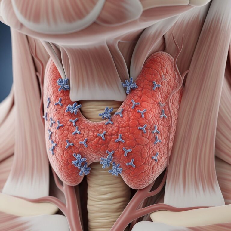
A schwannoma is a benign peripheral nerve sheath tumor that originates from Schwann cells, which are specialized cells that line and support peripheral nerves throughout the body. These tumors develop in the peripheral nervous system and grow slowly, making them non-cancerous and generally not an immediate health risk. Schwann cells function like insulation around nerve fibers, protecting them with a fatty substance called myelin that helps maintain proper nerve function and signal transmission. When these cells grow uncontrollably, they form a schwannoma.
Schwannomas are characterized by their well-circumscribed, encapsulated appearance with a consistent collagenous capsule that distinguishes them from other nerve sheath tumors. The tumor typically displaces the nerve root but does not envelope it, representing a key difference from neurofibromas. These tumors are considered the most common type of benign peripheral nerve tumor in adults and are more frequently observed in adults than in children.
Origin and Development
Schwannomas originate from malfunction and uncontrolled growth of Schwann cells that comprise the nerve sheath. The pathogenesis of schwannomas involves genetic alterations, particularly homozygous Nf2 loss in the Schwann cell lineage, which leads to schwannoma formation in experimental models. Unlike neurofibromas, which incorporate a mixture of non-neoplastic peripheral nerve components including axons, perineurial cells, fibroblasts, and inflammatory elements, schwannomas are composed primarily of neoplastic Schwann cells.
Most schwannomas are solitary tumors that occur in isolation without association with systemic genetic problems, termed “sporadic” schwannomas. However, multiple schwannomas can develop in patients with Neurofibromatosis Type 2 (NF2), a genetic condition in which patients are predisposed to multiple schwannomas throughout their bodies and other brain tumors. Additionally, schwannomas can be associated with a condition called schwannomatosis, which involves the development of multiple non-cutaneous neurofibromas and schwannomas.
Pathological Characteristics
Histologically, schwannomas display distinctive features that aid in their identification and diagnosis. The gross appearance is characteristically well-circumscribed with degenerative changes and variable admixture of compact spindled areas called Antoni A regions and hypocellular, microcystic Antoni B areas rich in macrophages and collagen fibers. A well-formed collagenous capsule and hyalinized vessels are consistent findings that distinguish schwannomas from other nerve sheath pathology.
By immunohistochemistry, schwannomas typically show diffuse, strong expression of S100 protein and abundant pericellular collagen type IV, consistent with the presence of a continuous pericellular basal lamina. These immunohistochemical markers are crucial for differential diagnosis, particularly distinguishing schwannomas from malignant peripheral nerve sheath tumors (MPNSTs), where diffuse S100 protein expression is exceedingly uncommon.
Cellular schwannomas represent a variant composed almost entirely of compact, fascicular proliferation of well-differentiated, cytologically bland Schwann cells. Despite their occasional alarming cellularity and higher local recurrence rates (5-40%) compared to conventional schwannomas, cellular schwannomas lack malignant potential for practical purposes and never metastasize.
Symptoms and Clinical Presentation
Schwannomas can cause various symptoms depending on their size, location, and the nerves they compress. Common symptoms include:
- Pain and discomfort at the tumor site or radiating pain along the affected nerve
- Numbness and tingling sensations (paresthesias) in the distribution of the affected nerve
- Weakness or loss of muscle function in areas supplied by the compressed nerve
- Balance issues and coordination problems, particularly with vestibular schwannomas
- Changes in hearing, especially with vestibular nerve involvement
- Visible swelling or mass under the skin in some cases
Symptoms may develop gradually as the tumor grows and exerts increasing pressure on adjacent neural structures. In many cases, particularly with small, slow-growing tumors, patients may experience no symptoms at all and the tumor is discovered incidentally during imaging for other reasons.
Common Locations
Schwannomas can develop throughout the body wherever peripheral nerves are present. A particularly common location is the vestibular nerve, which connects the brain to the inner ear; these tumors are called vestibular schwannomas (also known as acoustic neuromas). Other frequently affected sites include:
- The sciatic nerve in the leg
- The brachial plexus nerves in the arm and shoulder region
- The sacral plexus in the lower back
- Peripheral nerves of the extremities
- Spinal nerves and nerve roots
The distribution of schwannomas reflects the ubiquitous presence of Schwann cells throughout the peripheral nervous system, allowing these tumors to potentially develop anywhere peripheral nerves exist.
Diagnosis
Diagnosis of schwannomas typically involves a combination of clinical evaluation and imaging studies. Magnetic resonance imaging (MRI) is the gold standard imaging modality, providing excellent soft tissue contrast to visualize the tumor, assess its relationship to surrounding structures, and evaluate for associated features. MRI can demonstrate the characteristic well-circumscribed appearance of schwannomas and their displacement of adjacent neural structures.
Computed tomography (CT) may be used as an adjunctive imaging tool, particularly to assess bony involvement if the tumor is located near the spine or skull base. In some cases, electromyography (EMG) and nerve conduction studies may be performed to evaluate nerve function and document any neurological deficits prior to treatment planning.
While imaging characteristics are often diagnostic, histopathological examination through biopsy or examination of surgically resected tissue may be necessary to confirm the diagnosis and exclude malignant transformation, particularly if atypical features are present on imaging.
Risk of Malignant Transformation
Schwannomas are benign tumors that grow slowly and very rarely become malignant. However, rare cases of malignant transformation have been documented, resulting in malignant peripheral nerve sheath tumors (MPNSTs). Malignant schwannomas are more aggressive than their benign counterparts and can develop anywhere in the body, with particular predilection for the sciatic nerve of the leg, the brachial plexus nerves in the arm, and the sacral plexus in the lower back.
Malignant transformation is more commonly associated with neurofibromatosis type 1 (NF1), where these tumors may arise from preexisting benign neurofibromas that undergo malignant change. The prognosis for MPNSTs is challenging, as these tumors tend to be more aggressive with higher risk of recurrence and metastasis compared to benign schwannomas. Treatment typically involves a combination of surgery, radiation therapy, and chemotherapy aimed at removing the tumor and controlling its spread.
Treatment Options
Observation
For small, asymptomatic schwannomas or those growing slowly without causing neurological deficits, observation with periodic imaging follow-up may be appropriate. This conservative approach allows clinicians to monitor tumor growth and identify any changes that might warrant intervention. Many patients with asymptomatic schwannomas can be safely observed for years without requiring treatment.
Surgical Removal
Surgery to remove a schwannoma usually provides a cure and represents the primary treatment for symptomatic or enlarging tumors. Surgical resection aims to achieve complete tumor removal while preserving nerve function to the extent possible. The approach and technique vary depending on tumor location, size, and relationship to surrounding structures.
Neurosurgical expertise is essential for optimal outcomes, as careful dissection can often spare the parent nerve from significant injury. In many cases, particularly with smaller tumors or those with clear planes of separation from the nerve, complete tumor removal with preservation of nerve function can be achieved. Recovery from surgery varies depending on the extent of the procedure and the nerve involved, but most patients experience improvement in symptoms following successful resection.
Radiation Therapy
Radiation therapy may be considered in selected cases where surgical resection is not feasible or has been incomplete, or in patients with evidence of tumor recurrence or malignant transformation. Stereotactic radiosurgery, which delivers focused high-dose radiation to the tumor while minimizing exposure to surrounding tissues, may be employed for appropriately selected lesions. The role of radiation therapy continues to evolve as newer techniques become available.
Differential Diagnosis
Schwannomas must be differentiated from other nerve sheath tumors, most notably neurofibromas. While both are benign nerve sheath tumors, they differ in several important aspects:
| Feature | Schwannoma | Neurofibroma |
|---|---|---|
| Cell of Origin | Schwann cells (primary component) | Schwann cells with mixed non-neoplastic components |
| Relationship to Nerve | Displaces nerve, does not envelope | May envelope and incorporate nerve fibers |
| Genetic Association | Neurofibromatosis Type 2 (NF2) | Neurofibromatosis Type 1 (NF1) |
| Encapsulation | Well-defined collagenous capsule | Less consistently encapsulated |
| S100 Expression | Diffuse, strong | Variable |
| Malignant Potential | Extremely rare | Higher risk of transformation |
Hybrid tumors containing both schwannoma and neurofibroma components, or schwannoma and perineurioma components, have been documented and may present diagnostic challenges. Careful histopathological examination with immunohistochemical studies is often necessary to accurately classify these tumors and guide appropriate management.
Prognosis
The prognosis for benign schwannomas is generally excellent. These tumors grow slowly and rarely cause life-threatening complications. Complete surgical resection typically results in cure, with recurrence rates being uncommon. Even when recurrence occurs, the tumors generally remain benign and can be managed with additional surgical intervention if necessary.
Quality of life following treatment depends on several factors, including the size and location of the tumor, the extent of nerve compression at presentation, and the success of surgical resection in preserving nerve function. Many patients experience improvement or resolution of symptoms after successful surgery, though some degree of persistent neurological deficit may remain if significant nerve damage had occurred before treatment.
Frequently Asked Questions
Q: How common are schwannomas?
A: Schwannomas are not common tumors overall, but they are the most common type of benign peripheral nerve tumor in adults. They are seen more frequently in adults than in children.
Q: Can schwannomas be cancerous?
A: Schwannomas are benign tumors that very rarely become malignant. While malignant peripheral nerve sheath tumors can develop from schwannomas, this is an uncommon occurrence.
Q: Is surgery always necessary for schwannomas?
A: No, surgery is not always necessary. Asymptomatic or slowly growing schwannomas may be monitored with periodic imaging. Surgery is typically recommended for symptomatic tumors, those causing progressive neurological deficits, or rapidly growing lesions.
Q: What is a vestibular schwannoma?
A: A vestibular schwannoma is a schwannoma that develops on the vestibular nerve, which connects the brain to the inner ear. This is a common location for schwannomas and may cause hearing changes, balance problems, and tinnitus.
Q: What is the recovery like after schwannoma surgery?
A: Recovery varies depending on tumor location, size, and the surgical approach used. Most patients can return to normal activities within several weeks to a few months. Physical rehabilitation may be helpful for some patients.
Q: Are schwannomas hereditary?
A: Most schwannomas are sporadic and not hereditary. However, schwannomas can be associated with genetic conditions like Neurofibromatosis Type 2 (NF2) or schwannomatosis, which involve increased risk of developing multiple schwannomas.
References
- Pathology of Peripheral Nerve Sheath Tumors — National Center for Biotechnology Information (NCBI), National Institutes of Health. 2013. https://www.ncbi.nlm.nih.gov/pmc/articles/PMC3629555/
- Benign Nerve Sheath Tumor: Schwannoma vs. Neurofibroma — Neurosurgeons of New Jersey. https://www.neurosurgeonsofnewjersey.com/blog/benign-nerve-sheath-tumor/
- Schwannoma: What It Is, Causes, Symptoms & Treatment — Cleveland Clinic. https://my.clevelandclinic.org/health/diseases/17877-schwannoma
- Schwannoma – Symptoms, Causes, Treatments — Barrow Neurological Institute. https://www.barrowneuro.org/condition/schwannoma/
- Schwannoma | Other Conditions — Cancer Research UK. https://www.cancerresearchuk.org/about-cancer/other-conditions/schwannoma
- Schwannoma – Overview — Mayo Clinic. https://www.mayoclinic.org/diseases-conditions/schwannoma/cdc-20352974
Similar Articles


Read full bio of Sneha Tete