Sclerema Neonatorum Pathology: Clinical And Microscopic Guide
Detailed histopathological analysis of sclerema neonatorum, a rare and severe panniculitis in critically ill preterm neonates.

Sclerema neonatorum is a rare and grave form of panniculitis primarily affecting critically ill preterm neonates, characterized by progressive hardening of the skin and subcutaneous tissues due to unique histopathological changes in adipose tissue.
Introduction
Sclerema neonatorum (SN) represents an extreme manifestation of subcutaneous fat pathology in newborns, typically emerging in the first week of life among gravely ill, preterm infants. This condition leads to diffuse induration where the skin becomes tightly bound to underlying muscle and bone, often signaling a poor prognosis. Unlike more benign panniculitides, SN involves minimal inflammatory response and is associated with high mortality rates, frequently linked to underlying sepsis, hypothermia, or congenital anomalies.
The pathology is distinguished by the crystallization of lipids within adipocytes, resulting in the clinical picture of waxy, non-pitting hardening. This article delves into the microscopic features, clinical correlations, differential diagnoses, and investigative approaches, drawing from peer-reviewed case studies and dermatopathological expertise.
Clinical and Demographic Features
SN predominantly strikes preterm or low-birth-weight neonates who are already compromised by serious illnesses such as sepsis, respiratory distress, or gastrointestinal disorders. Onset is usually within the first seven days, though it may appear later in term infants under extreme stress. The process initiates in areas rich in subcutaneous fat, such as the buttocks, thighs, or trunk, sparing palms, soles, face, and genitalia.
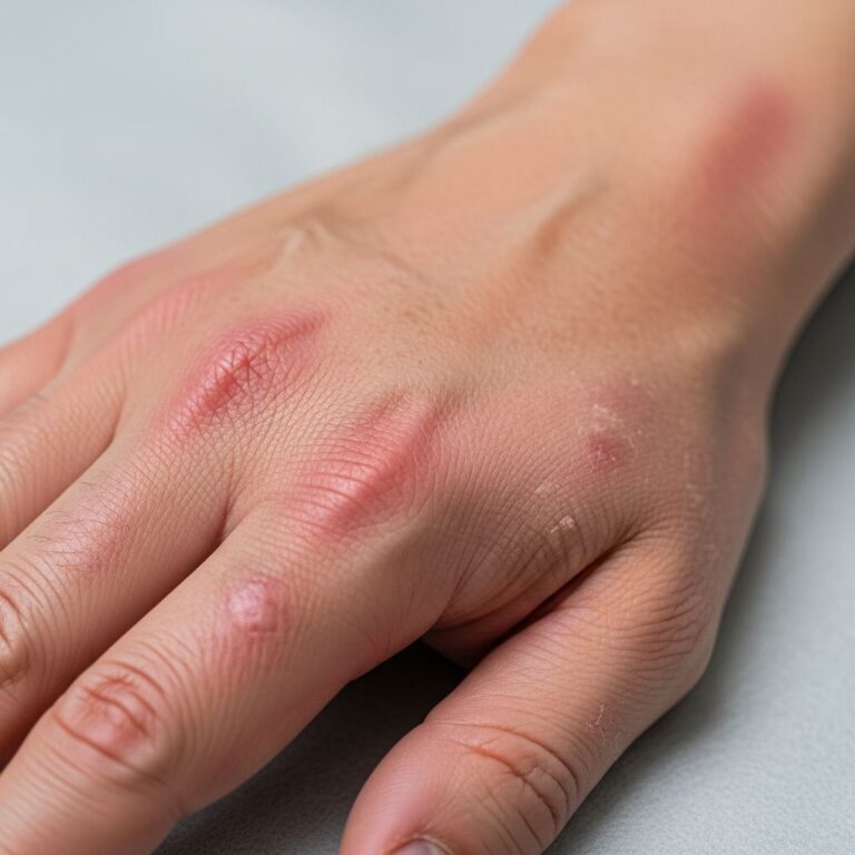
Affected skin appears smooth, cool, tense, mottled, purplish, and indurated, with no pitting on pressure. Progression is rapid, involving the trunk and extremities within 48 hours, compromising mobility, respiration, and feeding. In a documented term infant case, diffuse hardening began on the lower back on day 2, sparing spared areas, with normal vital signs initially but ultimate fatal outcome.
- Key demographics: Preterm (<37 weeks), low birth weight (<2500g), critically ill.
- Onset: First week of life, rapid spread.
- Spares: Palms, soles, genitalia, face.
- Prognosis: Mortality >50%, often due to underlying disease.
Microscopic Features
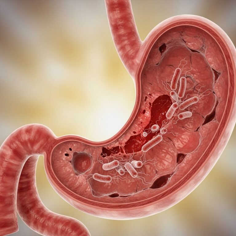
Histopathological examination is pivotal for definitive diagnosis, revealing hallmark needle-shaped clefts or crystals arranged radially within adipocytes. These represent crystallized saturated fats, a consequence of hypothermia or tissue hypoxia.
The subcutaneous fat shows thickened trabeculae with sparse lymphocytic and histiocytic infiltration, lacking significant fat necrosis or foreign body giant cell reaction. Dermal changes include dense collagen deposition, epidermal atrophy, and entrapment of adnexal structures. No edema or marked inflammation is present, distinguishing it from reactive panniculitides.
Low power view: Lobular panniculitis with sclerotic adipose septa binding skin to deeper tissues.
High power view: Radial needle-like spaces in adipocytes (cholesterol clefts), minimal inflammatory cells.
| Feature | Description |
|---|---|
| Adipocytes | Radially oriented needle-shaped crystals/clefts |
| Infiltrate | Sparse lymphocytes, histiocytes; no giant cells |
| Dermis | Thick collagen bundles, adnexal entrapment |
| Epidermis | Atrophy, reduced rete ridges |
| Other | No necrosis, no edema |
These findings confirm SN and exclude infectious or inflammatory mimics.
Pathophysiology
The exact mechanism remains obscure, but SN arises from altered subcutaneous fat metabolism in stressed neonates. Preterm fat is rich in saturated fatty acids, prone to solidification at lower temperatures, leading to crystal formation and vascular compromise. Hypoxia, hypothermia, and sepsis exacerbate this, causing fat crystallization without robust immune response due to neonatal immaturity.
Unlike subcutaneous fat necrosis of the newborn (SCFN), which shows inflammatory reaction around crystals, SN features acellular crystal deposition, reflecting terminal illness.
Differential Diagnosis
SN must be differentiated from other neonatal panniculitides and sclerotic conditions:
- Subcutaneous fat necrosis of the newborn (SCFN): Term/post-term infants; tender, erythematous nodules; crystals with mixed inflammatory infiltrate and fat necrosis.
- Scleredema neonatorum: Preterm; pitting edema, inflammatory edema on biopsy; self-limiting.
- Poststeroid panniculitis: History of steroids; similar crystals but with foreign body response.
- Infectious panniculitis: Bacterial/fungal; suppuration, organisms on special stains.
- Connective tissue diseases: Rare in neonates; more dermal involvement.
Clinical context and biopsy resolve most cases.
Investigation
Diagnosis is primarily clinical, supported by histopathology. No routine labs are specific, but sepsis workup (cultures, CRP) is essential. Skin biopsy (punch, 4-6mm) from indurated area is gold standard, avoiding cosmetically sensitive sites.
Imaging (X-ray) rules out calcifications or skeletal issues; normal in pure SN.
Treatment and Outcome
No specific therapy exists; management targets underlying illness (antibiotics for sepsis, warming). Experimental approaches like exchange transfusion or steroids lack evidence and are risky. Prognosis is dismal, with mortality from comorbidities rather than SN itself. Survivors rare, with resolution over weeks.
Frequently Asked Questions (FAQs)
What is sclerema neonatorum?
A rare panniculitis in sick preterm neonates causing skin hardening due to fat crystal formation.
How does it differ from SCFN on pathology?
SN has minimal inflammation around radial fat crystals; SCFN shows lobular panniculitis with giant cells.
Is biopsy always needed?
Clinical features are characteristic, but biopsy confirms and excludes differentials.
What is the prognosis?
High mortality (>50%) due to underlying critical illness.
Can term infants get SN?
Yes, though rare; often with sepsis or anomalies.
Images in Pathology
(Description: Low-power view shows sclerotic subcutaneous septa with bound-down dermis. High-power reveals adipocytes packed with radial needle-shaped clefts, sparse lymphocytes.)
References
- Sclerema Neonatorum in a Term Infant: A Case Report and Literature Review — Case Rep Dermatol. 2021. https://pmc.ncbi.nlm.nih.gov/articles/PMC7790589/
- Subcutaneous fat necrosis of the newborn — DermNet NZ. 2023. https://dermnetnz.org/topics/subcutaneous-fat-necrosis-of-the-newborn
- Subcutaneous fat necrosis of the newborn pathology — DermNet NZ. 2015. https://dermnetnz.org/topics/subcutaneous-fat-necrosis-of-the-newborn-pathology
- Sclerema neonatorum — DermNet NZ. 2023. https://dermnetnz.org/topics/sclerema-neonatorum
- Sclerema neonatorum pathology — DermNet NZ. 2015. https://dermnetnz.org/topics/sclerema-neonatorum-pathology
- Sclerema neonatorum – DermNet PRO — DermNet PRO. 2023. https://pro.dermnetnz.org/dermal-infiltrative/sclerema-neonatorum.html
Similar Articles


Read full bio of Sneha Tete