Sebopsoriasis: Expert Guide To Symptoms, Causes, And Treatment
Understanding sebopsoriasis: the overlap of seborrhoeic dermatitis and psoriasis, with causes, symptoms, diagnosis, and effective treatments.

Sebopsoriasis, also known as sebopsoriasis or seborrhoeic psoriasis, is a chronic inflammatory skin condition that combines clinical features of both seborrhoeic dermatitis and psoriasis. It typically affects sebaceous gland-rich areas such as the scalp, face, ears, chest, and flexures, presenting with well-defined erythematous plaques covered by greasy yellow scales. This hybrid disorder is more common in adults with a genetic predisposition to psoriasis and can be challenging to distinguish from its parent conditions.
What is sebopsoriasis?
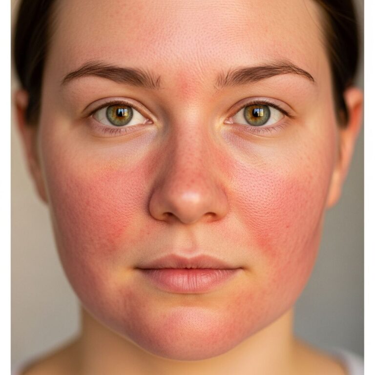
Sebopsoriasis represents an overlap syndrome where the flaky, itchy rashes of seborrhoeic dermatitis merge with the thicker, more defined plaques of psoriasis. Unlike classic psoriasis, which favours extensor surfaces, sebopsoriasis localises to seborrhoeic zones including the nasolabial folds, eyebrows, postauricular skin, and upper chest. The condition is relapsing-remitting, often triggered by stress, cold weather, or immune dysregulation, and may progress to full-blown psoriasis in some patients.
Authoritative sources describe it as a variant occurring in psoriasis-prone individuals who develop seborrhoeic dermatitis-like eruptions. The term ‘sebopsoriasis’ highlights this diagnostic ambiguity, as histopathology may show features of both disorders, such as parakeratosis, spongiosis, and Malassezia yeast proliferation. Prevalence is higher in males and those with HIV or Parkinson’s disease, where immune compromise exacerbates seborrhoeic involvement.
Who gets sebopsoriasis?
Sebopsoriasis primarily affects adults aged 20-50 years, with a male predominance due to higher sebaceous gland activity. Individuals with a family history of psoriasis are at elevated risk, as genetic factors like HLA-Cw6 alleles predispose to psoriatic eruptions in seborrhoeic areas. Comorbidities such as obesity, metabolic syndrome, and neurological disorders (e.g., Parkinson’s) increase susceptibility, likely through altered immune responses and sebum overproduction.
- Genetic predisposition: Up to 30% of first-degree relatives of psoriasis patients may develop sebopsoriasis.
- Immunocompromised states: HIV patients show a 5-10 fold higher incidence.
- Demographics: More common in fair-skinned individuals but can affect all skin types.
Infants rarely exhibit true sebopsoriasis, though cradle cap (infantile seborrhoeic dermatitis) may mimic it. The condition is not contagious and stems from endogenous factors rather than external pathogens.
What causes sebopsoriasis?
The aetiology of sebopsoriasis is multifactorial, blending psoriasis’s autoimmune pathology with seborrhoeic dermatitis’s microbial triggers. Central to both is an abnormal immune response to Malassezia yeast species (formerly Pityrosporum), which thrive in lipid-rich seborrhoeic areas. In genetically susceptible hosts, Malassezia lipases provoke T-cell activation, cytokine release (IL-17, IL-23), and keratinocyte hyperproliferation, mimicking psoriatic inflammation.
Key contributing factors include:
- Microbial overgrowth: Malassezia furfur proliferation due to excess sebum.
- Immune dysregulation: Defective skin barrier and Th17-mediated inflammation.
- Environmental triggers: Stress, cold dry weather, harsh soaps.
Unlike pure seborrhoeic dermatitis, sebopsoriasis involves stronger psoriatic genetic markers. Psoriasis genome-wide association studies identify risk loci like PSORS1 that may drive plaque formation in seborrhoeic zones.
What are the clinical features of sebopsoriasis?
Lesions appear as erythematous plaques with adherent, yellowish, greasy scales in a ‘sebaceous’ distribution. Common sites include:
- Scalp: Diffuse scaling extending beyond the hairline, resembling severe dandruff.
- Face: Eyebrows, glabella, nasolabial folds, ears with petaloid or annular patterns.
- Upper trunk: Presternal rhomboidal plaques.
- Flexures: Axillae, groin with less pronounced scaling.
Symptoms range from mild pruritus to intense itch, with potential for secondary bacterial infection if excoriated. Unlike classic psoriasis, Auspitz sign (pinpoint bleeding) is rare, but nail pitting may occur in 20-30% of cases. Darker skin types show more violaceous hue with finer scales.
| Feature | Sebopsoriasis | Seborrhoeic Dermatitis | Psoriasis |
|---|---|---|---|
| Scale | Greasy, yellow | Greasy, branny | Dry, silver |
| Distribution | Seborrhoeic areas | Seborrhoeic areas | Extensor surfaces |
| Thickness | Moderate plaque | Thin | Thick |
Diagnosis
Diagnosis is primarily clinical, based on characteristic distribution and morphology. Differential includes seborrhoeic dermatitis, psoriasis, tinea capitis, and contact dermatitis. Dermoscopy reveals ‘comma hairs’ from Malassezia and yellow clods. If ambiguity persists, skin biopsy shows psoriasiform hyperplasia with spongiosis and yeast forms.
Wood’s lamp may highlight Malassezia (coral pink fluorescence), and fungal KOH prep confirms hyphae. Blood tests rule out systemic psoriasis or HIV.
Treatment of sebopsoriasis
Treatment mirrors both parent conditions: antifungal agents control yeast, while antipsoriatics address hyperproliferation. A stepwise approach starts with topicals, escalating to systemic therapies for recalcitrant cases.
General measures
- Mild cleansers, emollients to reduce irritation.
- Avoid triggers: alcohol, stress, harsh hair products.
Scalp treatment
Medicated shampoos are first-line, used 2-3 times weekly:
- Ketoconazole 2% or ciclopirox (antifungal).
- Coal tar or salicylic acid for keratolysis.
- Steroid shampoos (clobetasol) for inflammation.
Facial and body treatment
- Antifungals: Ketoconazole cream 2% BD for 4 weeks.
- Topical steroids: Mometasone 0.1% OD, potent but short-term to avoid atrophy.
- Calcineurin inhibitors: Tacrolimus 0.1% or pimecrolimus for steroid-sparing, especially face.
Moderate-severe disease
- Phototherapy: Narrowband UVB effective for scalp/trunk.
- Systemic: Methotrexate, acitretin, or biologics (ixekizumab, guselkumab) if extensive[10].
Maintenance with antifungals prevents relapse. HIV-associated cases require antiretroviral therapy.
What is the outcome for sebopsoriasis?
Sebopsoriasis is chronic but manageable; 70% achieve clearance with combined therapy, though relapse is common upon discontinuation. Progression to guttate or plaque psoriasis occurs in 10-20%. Regular dermatologist follow-up optimises control and monitors for complications like tachyphylaxis or skin atrophy.
Frequently Asked Questions (FAQs)
Is sebopsoriasis the same as dandruff?
No, dandruff is mild seborrhoeic dermatitis; sebopsoriasis features thicker plaques with psoriatic elements.
Can sebopsoriasis be cured?
No cure exists, but symptoms remit with treatment in most cases.
Does diet affect sebopsoriasis?
Anti-inflammatory diets may help; avoid alcohol and gluten if sensitive.
Is sebopsoriasis contagious?
No, it is not infectious despite Malassezia involvement.
References
- Seborrheic Dermatitis: Causes, Symptoms, Treatment — National Eczema Association. 2023. https://nationaleczema.org/types-of-eczema/seborrheic-dermatitis/
- Scalp Psoriasis: Symptoms, Plaque, Causes & Treatment — Cleveland Clinic. 2024-01-15. https://my.clevelandclinic.org/health/diseases/22828-scalp-psoriasis
- Understanding Psoriasis – Causes, Symptoms & Treatment Options — Northeast Georgia Dermatology. 2023-05-10. https://negeorgiaderm.com/understanding-psoriasis-causes-symptoms-treatment-options/
- Scalp Psoriasis: Symptoms and Treatment — Dermatology OK. 2023. https://www.dermatologyok.com/blog/230551-scalp-psoriasis-symptoms-and-treatment
- Sebopsoriasis – DermNet — DermNet NZ. 2024. https://dermnetnz.org/topics/sebopsoriasis
- Scalp psoriasis vs. seborrheic dermatitis: What’s the difference? — Mayo Clinic. 2023-11-20. https://www.mayoclinic.org/diseases-conditions/psoriasis/expert-answers/scalp-psoriasis/faq-20058544
Similar Articles


Read full bio of medha deb