Sister Mary Joseph Nodule: 4 Causes, Key Signs, Diagnosis
Umbilical metastasis signaling advanced cancer: clinical features, diagnosis, and dire prognosis explained.

This eponymous term refers to secondary cancer (metastasis) appearing in the umbilicus, usually spreading from advanced peritoneal adenocarcinoma. The
Sister Mary Joseph nodule
(SMJN) is a rare but significant clinical sign that often heralds disseminated malignancy, primarily from gastrointestinal or gynecological primaries.What is the Sister Mary Joseph nodule?
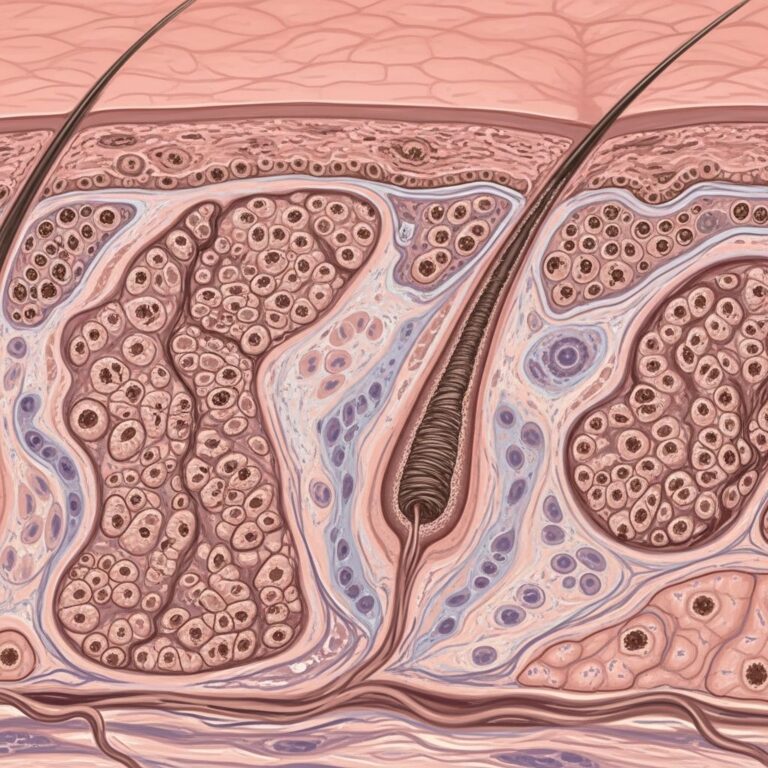
The Sister Mary Joseph nodule is a palpable, often protruding lesion at the umbilicus representing metastatic cancer from an intra-abdominal or pelvic primary tumor. It typically manifests as a firm, irregular nodule measuring 0.5 to 2 cm in diameter, which may ulcerate, become necrotic, or exude pus, blood, or serous fluid. This nodule is encountered in 1% to 3% of patients with malignant neoplasms of the gastrointestinal tract or ovary.
Clinically, it presents as a hard, fixed nodule that can be painful, with surface changes like oozing (50%) or ulceration (36.7%). Satellite lesions nearby occur in about 26.7% of cases, and the color is predominantly erythematous or red (66.7%). Dermoscopically, SMJN shows a characteristic polymorphous vascular pattern with white or milky-red amorphous areas, indicative of neoangiogenesis in malignant growth.
Who is Sister Mary Joseph?
Sister Mary Joseph Dempsey (1856–1939) was a Catholic nun and surgical assistant to Dr. William Mayo at St. Mary’s Hospital in Rochester, Minnesota. She noticed that patients with a palpable umbilical nodule during abdominal surgery had a poor prognosis. This observation led to the eponymous naming of the lesion as the “Sister Mary Joseph nodule”. Her keen clinical insight highlighted the prognostic significance of this finding long before modern imaging techniques.
What causes the Sister Mary Joseph nodule?
The nodule arises from metastasis to the umbilicus via several routes:
- Lymphatic spread: Through paraumbilical skin lymphatics draining to internal mammary nodes.
- Vascular embolization: Via portal vein branches connected to the umbilical vein remnant.
- Direct extension: From peritoneal carcinomatosis through the urachus or falciform ligament.
- Retrograde lymphatic spread: From involved liver hilum.
In approximately 15% of cases, no primary tumor is identified despite thorough investigation. The urachus and ligamentum teres provide potential pathways for tumor seeding to the umbilicus.
What are the most common primary malignancies?
Most patients (about 70-80%) have gastrointestinal malignancies, with the following distribution:
| Primary Site | Frequency |
|---|---|
| Gastric | ~30-50% |
| Colonic | ~25% |
| Pancreatic | ~20% |
| Ovarian/Uterine | ~15-20% |
| Other (breast, lung, unknown) | ~15% |
Gastric adenocarcinoma is the most frequent primary, followed by colorectal and pancreatic cancers. Gynecological malignancies, particularly ovarian carcinoma, account for a significant portion in females. Rarely, primaries from breast, lung, or unknown sites are reported.
Clinical features
Patients typically present with an asymptomatic or mildly irritating umbilical nodule. Key features include:
- Firm, irregular nodule (0.5-15 cm diameter).
- Erythematous, hard consistency with fixed base.
- May be painful; often ulcerated or discharging serous fluid, pus, or blood.
- Associated signs: ascites, pleural effusion, weight loss, or poor general state.
- In 15% of cases, it may be the only presenting sign in otherwise well patients.
An illustrative case involves a 70-year-old woman with umbilical irritation, papules, and patchy erythematous skin nearby (lymphangitis carcinomatosa). Imaging like CT or PET often reveals the primary tumor with peritoneal/liver metastases.
Differential diagnosis
SMJN must be distinguished from benign and malignant umbilical lesions. Clinical and dermoscopic features aid differentiation:
| Lesion Type | Clinical Features | Dermoscopic Features |
|---|---|---|
| Sister Mary Joseph Nodule | Red, oozing/ulcerated, satellites, hard | Polymorphous vessels, milky-red/white areas |
| Epidermal cyst | Blue, smooth, skin-toned | “Pore” sign |
| Dermatofibroma | Skin-toned, firm | Pigment network + white area |
| Verruca | Verrucous surface | Thrombosed capillaries |
| Epidermal nevus | Black, verrucous | Diffuse pigmentation |
| Keloid | Dusky red, scarred | Non-specific |
| Other | Pyogenic granuloma, hernia, endometriosis | Other vascular/inflammatory patterns |
Benign lesions often show specific dermoscopic patterns matching their histology, unlike the malignant vascular chaos of SMJN.
Investigations
Skin biopsy of the nodule is essential for diagnosis. Histology typically reveals adenocarcinoma with signet-ring cells in gastric primaries. Immunohistochemistry aids origin identification:
- CK7+/CK20+ for colorectal (CDX-2+).
- CEA, CA19-9 tumor markers.
- Imaging: CT/PET for primary and staging.
Biopsy confirmation prompted systemic staging in reported cases, revealing peritoneal and hepatic metastases.
Management
Treatment is palliative due to advanced disease:
- Chemotherapy: Systemic for primary tumor control.
- Surgical debulking: Rarely feasible if localized.
- Symptom management: Paracentesis for ascites, wound care.
- Palliative care: Focus on quality of life.
Early recognition via dermoscopy and biopsy enables prompt oncology referral.
Prognosis
The prognosis is extremely poor. Most patients succumb within 1 year due to widespread abdominal metastases. SMJN indicates stage IV disease with peritoneal carcinomatosis. Survival ranges from weeks to months post-diagnosis.
Frequently Asked Questions
Q: Is Sister Mary Joseph nodule always cancerous?
A: Yes, by definition it represents metastatic malignancy. Benign mimics exist but lack malignant dermoscopic/histologic features.
Q: Can it be the first sign of cancer?
A: Yes, in ~15% of cases, no primary is found initially, and it may present in asymptomatic patients.
Q: What does dermoscopy show?
A: Polymorphous vascular pattern with milky-red/white amorphous areas, pathognomonic for malignancy.
Q: Which cancer is most common?
A: Gastric adenocarcinoma (30-50%), followed by colorectal and pancreatic.
Q: How is it treated?
A: Palliative chemotherapy and supportive care; curative intent is rare.
References
- Benign Umbilical Tumors Resembling Sister Mary Joseph Nodule — PMC/NCBI. 2021-04-07. https://pmc.ncbi.nlm.nih.gov/articles/PMC8013636/
- Dermoscopic Features of Sister Mary Joseph Nodule — Actas Dermo-Sifiliográficas. 2015-07-01. https://www.actasdermo.org/en-dermoscopic-features-sister-mary-joseph-articulo-S1578219015001341
- Sister Mary Joseph nodule of the umbilicus — DermNet NZ. 2023. https://dermnetnz.org/topics/sister-mary-joseph-nodule-of-the-umbilicus
- Image of the Month—Diagnosis: Sister Mary Joseph Nodule — JAMA Surgery. 2010-02-01. https://jamanetwork.com/journals/jamasurgery/fullarticle/406864
- Sister Mary Joseph’s nodule: an unusual but important physical sign — PMC/NCBI. 2013-10-01. https://pmc.ncbi.nlm.nih.gov/articles/PMC3782795/
Similar Articles


Read full bio of medha deb