Skin Manifestations of Gastrointestinal Disease
Explore the vital links between gastrointestinal disorders and skin signs, aiding early diagnosis and treatment.

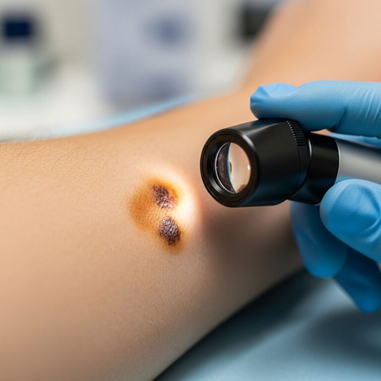
The skin and gastrointestinal (GI) tract share embryological origins from the ectoderm and endoderm, respectively, but exhibit profound clinical interconnections. Cutaneous manifestations often precede or accompany GI diseases, serving as critical diagnostic clues. Recognizing these skin signs enables early intervention, genetic screening for families, and multidisciplinary care involving dermatologists and gastroenterologists.
Who gets skin manifestations of gastrointestinal disease (and why)?
Skin manifestations arise in various GI disorders due to shared genetic, immunological, nutritional, or paraneoplastic mechanisms. They affect individuals with hereditary polyposis syndromes, inflammatory bowel disease (IBD), malabsorption states like celiac disease, and liver conditions. These signs may herald occult GI pathology, prompting investigation.
- Hereditary syndromes: Polyposis (e.g., Peutz-Jeghers, Gardner), hamartomatous polyps (e.g., Bannayan-Riley-Ruvalcaba).
- IBD: Crohn disease (CD), ulcerative colitis (UC) – reactive lesions like erythema nodosum (EN), pyoderma gangrenosum (PG).
- Malabsorption: Celiac disease – dermatitis herpetiformis.
- Liver disease: Cirrhosis – vascular signs, xanthomas.
Key dermatological signs
Distinctive skin findings map to specific GI pathologies:
| GI Condition | Key Skin Signs |
|---|---|
| Peutz-Jeghers syndrome | Hyperpigmented macules on lips, mucosa, perioral |
| Bannayan-Riley-Ruvalcaba | Genital hyperpigmentation, acrochordons, lipomas |
| Crohn disease | Erythema nodosum (4-6%), pyoderma gangrenosum |
| Ulcerative colitis | Pyoderma gangrenosum (2%), aphthous ulcers |
| Celiac disease | Dermatitis herpetiformis (intensely pruritic vesicles) |
| Ehlers-Danlos syndrome (vascular type) | Translucent skin, easy bruising, varicose veins |
Polyposis syndromes
Familial adenomatous polyposis (FAP) / Gardner syndrome
FAP involves hundreds of colorectal adenomas; Gardner variant adds extraintestinal features. Cutaneous signs include epidermoid cysts (up to 70% of cases), often multiple on face, scalp, extremities. Other findings: lipomas, desmoid tumors (firm plaques), fibromas, alopecia. Cysts appear in adolescence, preceding polyps.
Peutz-Jeghers syndrome (PJS)
PJS features hamartomatous polyps and mucocutaneous pigmentation. Hyperpigmented 1-5 mm macules on lips, buccal mucosa, periorbital, nose, genitalia, fingers/hands. Lesions precede GI symptoms, fade post-puberty (except oral). Increased cancer risk (GI, breast, pancreas). Histology: increased melanosomes.
Bannayan-Riley-Ruvalcaba syndrome (BRRS)
BRRS overlaps PTEN hamartoma syndromes with macrocephaly, hamartomas, lipomatosis. Specific skin sign: penile/vulvar hyperpigmentation. Others: genital lentigines, acanthosis nigricans-like facial lesions, acrochordons (neck, axilla, groin), vascular malformations, lipomas. Histology shows lentiginous hyperplasia.
Inflammatory bowel disease (IBD)
IBD encompasses Crohn disease (any GI segment, transmural) and ulcerative colitis (colon/rectum, mucosal). HLA-B27 links to UC; IBD1 gene to CD. Skin involvement in 10-20%, categorized as reactive, specific (granulomatous), or nutritional.
Specific lesions
- Cutaneous Crohn: Genital swelling, ulcers, fissures; oral aphthae, cobblestoning.
- Metastatic Crohn: Tender nodules/plaques on legs, thighs.
Reactive lesions
Erythema nodosum (EN)
Most common IBD skin sign (CD 4-6%, UC 3%). Painful erythematous nodules on shins, mirroring disease activity. Histology: septal panniculitis, lymphohistiocytic infiltrate. Resolves with IBD treatment; biopsy rarely needed.
Pyoderma gangrenosum (PG)
Seen in 3% IBD (UC 5-12%, CD 1-2%), female predominance. Starts as pustule/plaque, rapid ulceration with violaceous undermined borders. Pathergy common. More prevalent in UC. Treat underlying IBD; steroids, biologics for skin.
Other IBD skin signs
- Aphthous ulcers (both UC/CD).
- Erythema multiforme, Sweet syndrome, leukocytoclastic vasculitis.
- Psoriasis, hidradenitis suppurativa (increased risk).
Cutaneous manifestations of celiac disease
Celiac disease (gluten enteropathy) links to dermatitis herpetiformis (DH) in 10-15%. Intensely pruritic symmetrical vesicles/papules on extensors (elbows, knees, buttocks, shoulders). Oral erosions possible. Due to IgA deposits; gluten-free diet + dapsone effective. Nail dystrophy (98%), alopecia, hyperpigmentation from malabsorption. Alopecia patchy to totalis; hyperpigmentation on face, palms, neck.
Other gastrointestinal diseases
Ehlers-Danlos syndrome (vascular type)
Connective tissue disorder with GI fragility. Skin: translucent, visible veins, easy bruising, delayed healing, keloids. Complications: arterial rupture, bowel perforation.
Autoimmune liver diseases
- Primary biliary cholangitis: Xanthomas (palmar crease, eyelids), xanthelasma, pruritus.
- Primary sclerosing cholangitis: Similar vascular signs.
Whipple disease
Tropheryma whipplei infection. Hyperpigmentation (face, extremities), alopecia; due to malabsorption.
Clinical approach
Skin signs warrant GI evaluation: family history, endoscopy, genetics (e.g., STK11 for PJS, APC for FAP). Multidisciplinary management key.
Frequently asked questions
What are the most common skin signs in IBD?
Erythema nodosum and pyoderma gangrenosum; EN more in CD, PG in UC.
Do skin lesions in polyposis syndromes fade?
In Peutz-Jeghers, perioral macules fade post-puberty; mucosal persist.
Is biopsy needed for erythema nodosum?
Usually clinical diagnosis; histology shows panniculitis if performed.
How is dermatitis herpetiformis treated?
Strict gluten-free diet lifelong; dapsone for symptoms.
Can skin signs predict GI cancer risk?
Yes, e.g., pigmentation in PJS/BRRS signals screening needs.
References
- Cutaneous manifestation of gastrointestinal disease — Rahvar M, et al. Journal of Gastrointestinal Oncology. 2016-04-01. https://jgo.amegroups.org/article/view/5110/html
- Cutaneous manifestations of gastrointestinal disease — PubMed Central. 2016-03-22. https://pmc.ncbi.nlm.nih.gov/articles/PMC4783618/
- Skin signs of gastrointestinal disease — DermNet NZ. 2023. https://dermnetnz.org/topics/skin-manifestations-of-gastrointestinal-disease
- Skin as a Reflection of Gut Health — Cureus Journal. 2024. https://pmc.ncbi.nlm.nih.gov/articles/PMC11552655/
- Skin as a Reflection of Gut Health: An Overview — Cureus. 2024-10-01. https://www.cureus.com/articles/302614-skin-as-a-reflection-of-gut-health-an-overview-of-dermatological-manifestations-in-primary-neoplastic-and-autoimmune-gastrointestinal-disorders
Similar Articles


Read full bio of medha deb