Skin Metastasis: Guide To Symptoms, Diagnosis & Treatment
Cutaneous metastases from internal cancers: clinical features, diagnosis, and management strategies for advanced disease.

Skin metastasis, also known as cutaneous metastasis (plural metastases), refers to the growth of cancer cells in the skin stemming from a primary internal malignancy. This condition typically arises late in the disease course after the primary cancer, such as breast or lung cancer, has been diagnosed. In rare instances, skin lesions may precede or coincide with the discovery of the internal tumour, prompting further investigation.
Cutaneous metastases can also originate from primary skin cancers, particularly melanoma, where the original tumour spreads to distant skin sites or other organs like the lungs or brain. The process involves cancer cells detaching from the primary tumour and travelling via the bloodstream or lymphatic system to lodge in the skin. While most malignant tumours can metastasise to the skin, certain cancers do so more frequently, with an overall incidence of 3–10% in patients with primary malignancies.
Who gets skin metastasis?
The likelihood of skin metastasis varies by cancer type, patient sex, and age. In women, approximately 70% of cases originate from breast cancer, reflecting its prevalence and propensity for lymphatic spread. In men, the most common sources are lung cancer (24%), colon cancer (19%), melanoma (13%), and oral cavity cancers (12%).
Age and sex influence patterns; for example, older adults may show higher rates from gastrointestinal cancers. The regional distribution often correlates with the primary tumour’s location: chest wall for breast cancer, anterior trunk for lung, and scalp or extremities for melanoma.
- Women: Breast (70%), followed by lung, colorectal, ovarian.
- Men: Lung (24%), colon (19%), melanoma (13%), oral cavity (12%).
- Children: Rare, often from sarcomas or neuroblastomas.
Autopsy studies indicate skin involvement in up to 9% of cancer patients, though clinical detection is lower at 0.7–4.4%.
Related cancers
Numerous internal malignancies can produce skin metastases. Common ones include:
- Breast carcinoma
- Lung carcinoma (non-small cell and small cell)
- Colorectal carcinoma
- Renal cell carcinoma
- Gastric carcinoma
- Head and neck squamous cell carcinoma
- Melanoma (primary skin cancer metastasis)
Less frequent but notable: ovarian, prostate, bladder, hepatocellular carcinoma, and sarcomas.
Clinical features of skin metastasis
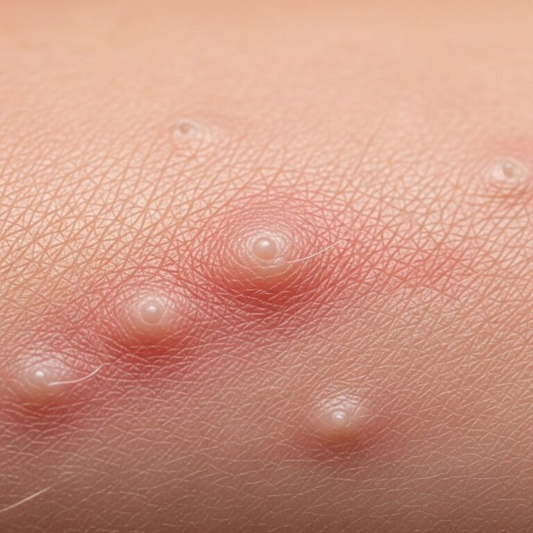
Skin metastases most commonly appear in regions adjacent to the primary tumour. The initial presentation is typically a firm, round or oval, mobile, non-painless nodule with a rubbery, firm, or hard texture. Sizes range from barely visible to several centimetres. Colours vary: skin-toned, red, violaceous, or black/blue in melanoma.
Multiple nodules may emerge rapidly, sometimes ulcerating or forming plaques. Specific patterns include:
- Carcinoma telangiectaticum: Telangiectasias (dilated blood vessels) over nodules.
- Carcinoma erysipeloides / carcinoma en cuirasse: Inflammatory, erysipelas-like erythema, often from breast cancer, leading to sclerotic ‘armour-like’ thickening.
- Carcinoma bullosum: Bullae overlying nodules.
- Carcinoma fistulosum: Fistulas draining fluid.
- Leonine facies: Facial thickening mimicking leonine facies in leprosy, from head/neck cancers.
- Zosteriform / lymphangitic: Dermatomal or linear lymphatic spread.
- Alopecia neoplastica: Scalp nodules causing alopecia.
Symptoms may include pain, pruritus, bleeding, or infection if ulcerated. Breast cancer skin metastases often affect the chest wall, presenting as nodules, inflammation resembling cellulitis, or lymphoedema.
Organ-specific features
| Organ of cancer origin | Features of cutaneous metastasis |
|---|---|
| Breast | Chest wall nodules, carcinoma en cuirasse (sclerotic plaque), telangiectatic carcinoma, inflammatory erysipelas-like patches. |
| Lung | Firm nodules on anterior chest; may mimic panniculitis or be zosteriform. |
| Melanoma | Amelanotic, pigmented, or ulcerated nodules; often multiple and widespread. |
| Colon and stomach | Scalp nodules (Sister Mary Joseph’s nodule for umbilicus in gastric ca); firm abdominal nodules. |
| Kidney | Vascular, red nodules (‘acquired vascular malformation’). |
| Ovary | Scalp nodules; umbilical Sister Mary Joseph’s nodule. |
| Prostate | Pea-sized perineal nodules; indurated plaques. |
[Adapted from DermNet NZ]
Diagnosis of skin metastasis
Suspicious skin lesions in cancer patients warrant biopsy. Histopathology reveals tumour cells resembling the primary malignancy, though dedifferentiation may occur. Immunohistochemistry (e.g., cytokeratins, melanoma markers like S100/HMB45) confirms origin.
Imaging (CT/PET) and primary tumour restaging are essential. Differential diagnoses include benign adnexal tumours, cysts, pyogenic granulomas, or infections. Early skin metastases may herald occult primaries, necessitating systemic workup.
Management of skin metastasis
Treatment targets the underlying primary cancer. When metastatic disease is widespread, systemic therapy (chemotherapy, targeted agents, immunotherapy) is prioritised, though often palliative.
For localised skin lesions:
- Surgical excision: Solitary nodules, if feasible.
- Radiotherapy: Painful/ulcerated lesions; effective for breast/lung metastases.
- Electrochemotherapy: Combines low-dose chemotherapy with electric pulses to enhance drug uptake in cancer cells; useful for cutaneous breast metastases.
- Topical therapies: Imiquimod, retinoids (experimental).
- Dressings/Debridement: For ulcerated, bleeding, or infected lesions to maintain hygiene and reduce odour.
- Palliative care: Pain management, antibiotics for secondary infection, wound care.
Prognosis is poor; skin metastases indicate advanced disease with median survival of 3–6 months, varying by primary (better for renal cell, worse for lung). Multidisciplinary input from oncologists, dermatologists, and palliative teams is crucial.
Prevention
Preventing metastasis involves early detection and aggressive treatment of primary cancers. Regular skin exams in high-risk patients (e.g., advanced breast/lung cancer) aid early intervention. UV protection reduces melanoma risk.
FAQ
What is skin metastasis?
A: The spread of cancer cells from an internal primary tumour to the skin via blood or lymphatics, appearing as nodules or plaques.
Which cancer most commonly metastasises to skin in women?
A: Breast cancer, accounting for ~70% of cases.
Are skin metastases painful?
A: Usually painless, but ulceration can cause pain, bleeding, or infection.
Can skin metastases be the first sign of cancer?
A: Yes, rarely; they may prompt discovery of an occult primary.
What is carcinoma en cuirasse?
A: Sclerotic, armour-like plaque on chest wall from breast cancer lymphatic spread.
Is treatment curative?
A: Rarely; focus is palliative once widespread.
References
- Skin metastasis, cutaneous metastases – DermNet — DermNet NZ. 2023-05-15. https://dermnetnz.org/topics/skin-metastasis
- Metastatic Melanoma: Causes, Symptoms, and Treatment — WebMD. 2024-08-20. https://www.webmd.com/melanoma-skin-cancer/metastatic-melanoma
- Skin metastasis: a pathologist’s perspective — Wiley Online Library (Journal of Cutaneous Pathology). 2009-11-01. https://onlinelibrary.wiley.com/doi/abs/10.1111/j.1600-0560.2009.01469.x
- Secondary (metastatic) breast cancer in the skin — Breast Cancer Now. 2024-03-10. https://breastcancernow.org/about-breast-cancer/secondary-breast-cancer/secondary-breast-cancer-treatment/secondary-breast-cancer-in-the-skin-skin-metastases
- Spread to the Skin — Say YES to HOPE. 2023-11-05. https://www.sayyestohope.org/spread-to-the-skin/
Similar Articles


Read full bio of medha deb