Skin Signs Of Neurological Diseases: 5 Key Associations To Know
Discover how skin changes can signal underlying neurological disorders and why early detection matters.

Skin Signs of Neurological Diseases
The relationship between skin and the nervous system is profound and bidirectional. The skin and central nervous system share common embryonic origins, originating from the neural crest, which gives rise to melanocytes and nerve tissues found throughout the brain and spinal cord. Because of this intimate connection, numerous neurological disorders manifest with visible skin changes that can serve as early warning signs or diagnostic indicators. Understanding these cutaneous manifestations is critical for both dermatologists and neurologists, as skin changes may occasionally be the first sign of an underlying neurological condition, potentially affecting disease prognosis and patient outcomes.
The Skin-Nervous System Connection
The skin is not merely a protective barrier; it functions as a complex neuroimmunoendocrine organ with extensive connections to the peripheral sensory nervous system, autonomic nervous system, and central nervous system. Dermatomes on the skin provide sensory information to the brain, while the skin contains numerous neuropeptides released from sensory nerves that play precise roles in cutaneous physiology and pathophysiology. When dysfunction occurs in the nervous system, these pathways often become dysregulated, resulting in visible skin manifestations that can alert healthcare providers to underlying neurological disease.
Parkinson’s Disease and Skin Manifestations
Parkinson’s disease presents multiple cutaneous manifestations, several of which may precede the development of classic motor symptoms by years. Recognizing these early warning signs is essential for timely diagnosis and intervention.
Seborrheic Dermatitis and Rosacea
Seborrheic dermatitis is among the most common skin conditions associated with Parkinson’s disease. This condition affects oily areas of the body, including the sides of the nose, eyebrows, ears, eyelids, and chest, causing scaly patches, inflamed skin, and dandruff. Rosacea, a chronic facial redness condition, has been identified as a potential early warning sign that may increase risk for Parkinson’s disease. While most people with rosacea do not develop Parkinson’s, studies demonstrate that rosacea often predates the development of tremors and other motor symptoms by several years.
Dry and Oily Skin Changes
Changes in skin texture and moisture regulation are common in Parkinson’s disease. Many people with PD develop oily or flaky skin, especially on the face and scalp. Conversely, extreme dryness of the skin can also be problematic, requiring the use of skin moisturizers and professional dermatological consultation.
Melanoma Risk
The most serious skin condition linked to Parkinson’s disease is melanoma. Many studies have demonstrated that patients with Parkinson’s have a significantly higher risk of developing melanoma. The mechanism behind this increased risk involves melanin dysregulation: people with Parkinson’s have less melanin in the brain due to degeneration of neurons that produce dopamine, which contains melanin. It is hypothesized that they may also have decreased melanin in the skin, reducing protective capacity against skin cancer. Notably, the relationship appears bidirectional—a 2017 Mayo Clinic study found that people with melanoma have four times the risk of developing Parkinson’s disease. Neurologists generally recommend that Parkinson’s patients receive annual dermatological skin checkups. Fortunately, recent studies have shown that levodopa, a primary Parkinson’s medication, is not associated with increased melanoma risk, allaying earlier concerns.
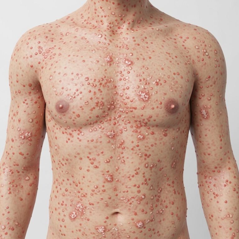
Bullous Pemphigoid and Neurological Disorders
Recent research has uncovered a significant bidirectional association between bullous pemphigoid and multiple neurological disorders. Bullous pemphigoid is a chronic blistering condition affecting the skin and mucous membranes. Studies published in the European Journal of Dermatology and other peer-reviewed journals have identified strong connections between bullous pemphigoid and neurological conditions including dementia, Parkinson’s disease, epilepsy/seizures, multiple sclerosis, and stroke.
The relationship is two-directional: people with these neurological conditions are more likely to develop bullous pemphigoid, and conversely, people who develop bullous pemphigoid are at increased risk for these neurological disorders. In one comprehensive study, 50.3 percent of patients with bullous pemphigoid had at least one neurologic disease prior to their blistering condition diagnosis, compared with only 24.2 percent in control groups. When a patient presents with a known neurologic disorder and a new blistering skin condition, bullous pemphigoid should be considered high on the diagnostic list.
Genetic Neurocutaneous Disorders
Genodermatoses—inherited genetic conditions affecting the skin and nervous system—frequently present with cutaneous manifestations that precede or accompany systemic neurological involvement. These conditions require expertise from neurologists, physicians, and dermatologists working collaboratively.
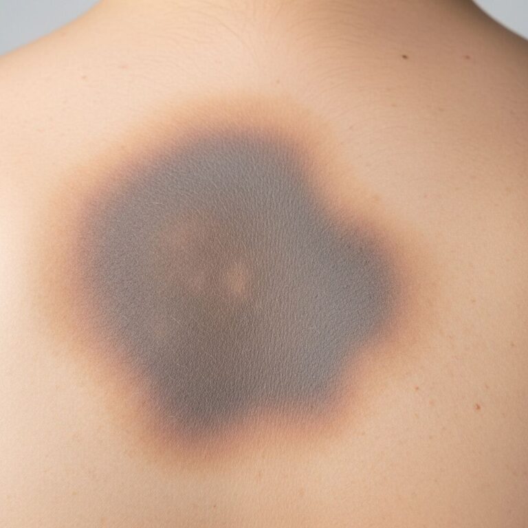
Tuberous Sclerosis Complex (TSC)
Tuberous sclerosis is characterized by hamartomas (benign tumors) affecting multiple organ systems, including the brain, skin, heart, and kidneys. Skin manifestations include ash leaf spots—hypopigmented lesions where the skin appears lighter than surrounding areas. When hypopigmented lesions are found in combination with seizures, they can indicate tuberous sclerosis or related conditions. Additional skin manifestations include facial angiofibromas, which are small, firm, smooth, dome-shaped raised spots typically pink to red in color, and café au lait macules—painless, light- to dark-brown-pigmented birthmarks.
Neurofibromatosis Type 1 (NF1)
Neurofibromatosis type 1 presents with multiple café au lait macules as a hallmark feature. While having one or two such spots is common in the general population, multiple café au lait spots can indicate neurofibromatosis and warrant neurological evaluation. The condition may also involve neurofibromas—benign tumors of nerve tissue—that manifest as raised bumps on or under the skin.
Other Genetic Conditions
Additional genetic neurocutaneous disorders include ataxia-telangiectasia, which presents with telangiectasias (dilated blood vessels visible as red marks on the skin) alongside progressive neurological symptoms. These genodermatoses may present cutaneous manifestations followed by systemic neurological manifestations, requiring careful clinical observation and multidisciplinary management.
Vasculitic Syndromes with Cutaneous and Neurological Involvement
Vasculitic syndromes—conditions involving inflammation of blood vessels—frequently present with simultaneous cutaneous and neurological involvement, possibly linked to common pathogenic mechanisms connecting the peripheral nervous system to the skin. These conditions include polyarteritis nodosa, Churg-Strauss syndrome, Wegener granulomatosis, and cryoglobulinemia, which commonly manifest with both skin and neurological signs.
Nerve Compression Syndromes with Skin Manifestations
Certain nerve compression conditions present with distinctive cutaneous involvement that may herald underlying neurological disease. Carpal tunnel syndrome, caused by median nerve compression, can present with sensation of itching, ulceration, blistering, hypohidrosis (decreased sweating), Raynaud’s phenomenon, and irritant contact dermatitis. Trigeminal trophic syndrome, involving trigeminal nerve pathology, presents with ala nasi ulceration and other distinctive cutaneous features.
These conditions illustrate an important clinical principle: dysregulation of neuropeptide release from peripheral nerve terminations secondary to compression creates an imbalance between immunostimulating and immunosuppressive neuropeptides, favoring the occurrence of dermatological signs. Dermatologists recognizing these skin changes should consider referral for neurological consultation to arrive at the correct underlying diagnosis.
Herpes Zoster and Neurological Sequelae
Herpes zoster (shingles) represents a classic example of a condition involving both cutaneous and nerve manifestations. The characteristic vesicular rash follows dermatome distribution, reflecting the pattern of viral reactivation along sensory nerve roots. Beyond the acute blistering phase, post-herpetic neuralgia—persistent neuropathic pain—can develop, representing significant neurological involvement requiring ongoing management.
Diagnostic Importance of Skin Examination
For any neurological condition, a thorough physical examination must include careful examination of the skin. There are literally hundreds of neurological disorders with manifestations in the skin or with skin-related side effects to their treatment. As one example, skin biopsy can serve as an aid to diagnosis of neurological disease and neuropathies, providing tissue evidence of neurological pathology.
A clinical scenario illustrates this principle: a child evaluated for suspicious birthmarks who subsequently experiences a seizure may present to a neurologist’s office. The combination of specific cutaneous findings with neurological symptoms can rapidly narrow the differential diagnosis, leading to appropriate testing and treatment.
Categories of Skin-Neurological Associations
Cutaneous manifestations of neurological disease can be categorized into several groups:
- Infections: Conditions like herpes zoster affecting both skin and peripheral nerves
- Metabolic diseases: Disorders affecting metabolism that present with both skin and neurological symptoms
- Connective tissue disorders: Systemic conditions like lupus with manifestations in both systems
- Genodermatoses: Inherited genetic conditions such as neurofibromatosis and tuberous sclerosis
- Nutritional deficiencies: Deficiencies affecting both skin health and neurological function
Clinical Implications for Dermatologists
Dermatologists play a critical role in identifying skin manifestations that may herald neurological disease. When encountering patients with syndromes of unclear etiology, dermatologists should consider whether neurological consultation is warranted. This is particularly important for patients presenting with cutaneous ulceration, blistering, unusual sensory symptoms, or progressive skin changes without clear dermatological etiology.
Protein Markers in Neurological Disease
Emerging research suggests that certain proteins associated with neurological disease may accumulate in skin tissue, potentially offering diagnostic insights. For example, in Parkinson’s disease, the alpha-synuclein protein typically accumulates only in the neck skin, whereas in multiple system atrophy, alpha-synuclein is more widely distributed in the skin. Progressive supranuclear palsy, which may initially resemble Parkinson’s clinically, involves tau protein accumulation but lacks alpha-synuclein in the skin. These protein signatures may eventually support diagnostic differentiation between conditions with overlapping clinical presentations.
Frequently Asked Questions
Q: Can skin conditions always indicate neurological disease?
A: Not all skin conditions indicate neurological disease. However, when skin manifestations appear in combination with neurological symptoms, or when specific patterns emerge (such as multiple café au lait spots or ash leaf spots), neurological evaluation becomes important.
Q: Should people with Parkinson’s disease have regular skin checks?
A: Yes. Due to the increased risk of melanoma in Parkinson’s disease, neurologists generally recommend that their patients see a dermatologist for annual skin checkups.
Q: How early can skin signs appear?
A: Some skin manifestations may precede neurological diagnosis by several years. For example, rosacea or seborrheic dermatitis may appear years before Parkinson’s disease symptoms develop.
Q: What should I do if I notice unusual skin changes?
A: Report any persistent or progressive skin changes to your primary care physician or dermatologist, who can determine whether neurological consultation is appropriate based on the specific findings.
Q: Are genetic skin-neurological conditions preventable?
A: Genetic neurocutaneous disorders like neurofibromatosis and tuberous sclerosis are inherited conditions that cannot be prevented, but early recognition through skin manifestations enables timely neurological evaluation and management.
References
- Skin Problems May Be Early Signs of a Neurologic Condition — Brain and Life Magazine. 2024. https://www.brainandlife.org/articles/skin-problems-early-signs-of-neurologic-condition
- Cutaneous Manifestations and Neurological Diseases — PubMed Central (PMC). 2024. https://pmc.ncbi.nlm.nih.gov/articles/PMC10642374/
- Dermatological and Immunological Conditions Due to Nerve Lesions — PubMed Central (PMC). 2013. https://pmc.ncbi.nlm.nih.gov/articles/PMC3812736/
- Skin Changes — Parkinson’s Foundation. 2024. https://www.parkinson.org/understanding-parkinsons/non-movement-symptoms/skin
- Neurological Conditions Affecting the Skin — Wiley Online Library. 2023. https://onlinelibrary.wiley.com/doi/10.1002/9781119709268.rook083
- Neurodermatitis: Symptoms and Causes — Mayo Clinic. 2024. https://www.mayoclinic.org/diseases-conditions/neurodermatitis/symptoms-causes/syc-20375634
Similar Articles


Read full bio of Sneha Tete