Skin Signs Of Coma: Clinical Guide To Diagnosis And Care
Recognizing cutaneous clues in comatose patients to diagnose underlying causes and manage complications effectively.

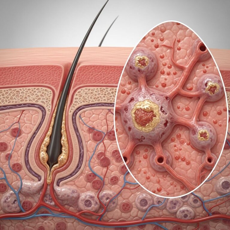
Coma represents a profound state of unconsciousness characterized by the absence of purposeful response to external stimuli, including pain, light, or sound. Patients in coma typically require intensive care unit (ICU) management due to the severity of their condition. Common precipitants include traumatic head injury, cerebrovascular accidents such as stroke, intracranial neoplasms like brain tumours, substance intoxication from drugs or alcohol, metabolic derangements in diabetes mellitus, and severe infections. A thorough dermatological examination in these patients can yield critical diagnostic insights into the aetiology of the coma or reveal secondary cutaneous changes resulting from the comatose state itself.
Introduction
The skin serves as a visible window into systemic pathology, particularly valuable in comatose individuals who cannot communicate symptoms. Cutaneous findings may precede neurological deterioration, offering clues to initiate targeted investigations and therapies. For instance, distinctive rashes, blisters, or pigmentation changes can point to specific infectious, toxic, metabolic, or vascular causes. Early recognition of these signs is pivotal, as it can expedite diagnosis and improve outcomes in time-sensitive conditions like bacterial meningitis or diabetic ketoacidosis.
Coma often stems from acute events such as serious falls, motor vehicle accidents, or assaults leading to head trauma. Chronic progressive diseases like malignancies or end-stage organ failure can also culminate in coma. Regardless of origin, immobility in coma predisposes patients to unique skin complications, including pressure-induced injuries and friction-related lesions.
Causes and Their Specific Skin Signs
Diverse aetiologies of coma manifest with characteristic cutaneous features, aiding in differential diagnosis. These signs can be categorized by underlying mechanism: infectious, toxic, vascular, metabolic/endocrine, nutritional, and paraneoplastic.
Infections
Coma frequently accompanies central nervous system (CNS) infections or systemic sepsis with encephalopathic features. Key examples include:
- Bacterial meningitis: Purpuric or petechial rash, often due to meningococcal septicaemia, progressing to purpura fulminans with necrotic lesions.
- Viral encephalitis: Maculopapular eruptions or vesicular rashes, as seen in herpes simplex virus encephalitis.
- Cryptococcal infection: Particularly in immunocompromised patients, presenting with molluscum-like umbilicated papules.
- Toxic shock syndrome and severe skin infections: Diffuse erythroderma, desquamation, and multi-organ failure with septic encephalopathy.
Peripheral clues like an attached tick may indicate tick-borne encephalitis, where the vector remains visible on the skin.
Toxins
Toxic ingestions or exposures frequently induce coma, with dermatological hallmarks:
- Cholinesterase inhibitor toxicity: Nicotinic signs such as miosis, fasciculations, and diaphoresis; organophosphate poisoning may show garlic-like odour and pinpoint pupils.
- Inhaled mercury exposure: Acrodynia with pink papules on acral sites, irritability, and hypertension.
Drug hypersensitivity reactions, including erythema multiforme or toxic epidermal necrolysis, can precipitate coma via severe systemic inflammation.
Vascular Causes
Cerebrovascular events are prime culprits in coma, distinguishable by embolic sources:
- Hypoxic-ischaemic encephalopathy: Often post-cardiac arrest, with mottled cyanosis.
- Embolic stroke: Cholesterol emboli present as livedo reticularis or blue toe syndrome; septic emboli cause pustular lesions; fat emboli from fractures yield petechiae on conjunctivae and axillae.
- Subarachnoid haemorrhage: Neurocutaneous syndromes like neurofibromatosis type 1 with café-au-lait macules.
- Cerebral vasculitis: Livedo reticularis from medium-vessel involvement, such as polyarteritis nodosa.
Seizures
Status epilepticus can lead to coma, with skin signs limited to trauma from falls or tongue-biting ecchymoses.
Systemic Diseases
Metabolic and endocrine derangements cause coma with telltale skin changes:
- Hepatic encephalopathy: Asterixis-induced excoriations, spider angiomata, palmar erythema, and jaundice.
- End-stage renal disease: Uraemic frost (urea crystals on skin), prurigo nodularis, and perforating disorders.
- Myxoedema coma: Non-pitting oedema, dry coarse skin, hair loss, and macroglossia in severe hypothyroidism.
- Hypoadrenalism (Addisonian crisis): Hyperpigmentation of buccal mucosa, creases, and gingival pigmentation as a diagnostic clue.
- Diabetic ketoacidosis: Bullosis diabeticorum (tense blisters on extremities), diabetic dermopathy (shin spots), or necrobiosis lipoidica.
Other Systemic Causes
Hyperviscosity syndrome from paraproteinaemias causes retinal haemorrhages and orocutaneous purpura. Cutaneous vasculitic lesions signal cerebral vasculitis.
Nutritional Deficiencies
Scurvy presents with perifollicular haemorrhages and corkscrew hairs, leading to coma from cerebral haemorrhage. Pellagra (niacin deficiency) shows photosensitive dermatitis, diarrhoea, and dementia.
Drug Hypersensitivity Syndromes
Severe cutaneous adverse reactions like Stevens-Johnson syndrome/toxic epidermal necrolysis feature mucosal erosions, epidermal sloughing, and Nikolsky sign, potentially causing coma from dehydration or sepsis.
Cutaneous Paraneoplastic Syndromes
Acanthosis nigricans, dermatomyositis (heliotrope rash, Gottron papules), or Sweet syndrome may herald underlying malignancies causing coma via metastases or hypercalcaemia.
Skin Signs from Being in a Coma
Prolonged immobility in coma engenders specific dermatoses due to pressure, shear, moisture, and nutritional deficits.
Pressure Ulcers
Also termed decubitus ulcers, these arise from sustained pressure over bony prominences (sacrum, heels, trochanters), compounded by hypotension, vasoactive drugs, malnutrition, and diabetes. Stages progress from non-blanchable erythema (Stage 1) to full-thickness necrosis exposing bone (Stage 4).
Prevention strategies:
- Frequent repositioning every 2 hours.
- Specialized pressure-redistributing mattresses.
- Moisture management and nutritional support.
- Correcting hypotension and glycaemic control.
Coma Blisters
These are multiple, tense, blood-filled or serous blisters at pressure points (knees, elbows, sacral area, feet), emerging 2–3 days post-coma onset. Historically linked to barbiturate overdose, they occur in any coma aetiology (e.g., diabetic, traumatic). Pathogenesis involves hypotension-induced epidermal necrosis, pressure ischaemia, and sweat gland/duct necrosis. Lesions are self-limiting, healing over weeks without scarring.
Histology reveals subepidermal clefting, sweat gland necrosis, and dermal vessel thrombi (suggesting non-drug aetiology). Differential includes bullous fixed drug eruption and porphyria.
Other Skin Signs
- Friction injuries: Abrasions from bed transfers.
- Moisture lesions: Intertrigo in dependent areas.
- Medical device-related pressure injuries: From oxygen masks, endotracheal tubes.
- Dependent lividity: Purple discolouration in recumbent positions.
Frequently Asked Questions (FAQs)
Q: What are coma blisters?
A: Tense, haemorrhagic blisters at pressure sites appearing 2–3 days into coma, due to necrosis from hypotension and pressure; they resolve spontaneously.
Q: How can skin examination help diagnose coma causes?
A: Specific rashes like purpura in meningitis, gingival pigmentation in Addison disease, or livedo in vasculitis provide aetiological clues.
Q: What preventive measures reduce pressure ulcers in coma?
A: Regular turning, pressure-relieving devices, nutrition, and comorbidity management like diabetes control.
Q: Are coma blisters always drug-induced?
A: No, biopsy showing vessel thrombi indicates non-drug causes; they occur in various comas.
Q: Which infections commonly cause coma with skin signs?
A: Bacterial meningitis (petechiae), viral encephalitis (vesicles), cryptococcosis (papules), and toxic shock (erythroderma).
Clinical Images and Descriptions
Visual aids enhance recognition:
- Gingival hyperpigmentation signalling Addisonian crisis.
- Diabetic bullae and shin dermopathy in hyperglycaemic coma.
- Temporal ulcer from giant cell arteritis.
- Livedo reticularis in cerebral vasculitis.
- Tick attachment in encephalitis.
References
- Skin signs of coma — DermNet NZ. 2022. https://dermnetnz.org/topics/skin-signs-of-coma
- Skin signs of coma images — DermNet NZ. 2022. https://dermnetnz.org/images/skin-signs-of-coma-images
- Dermatologic emergencies — National Institutes of Health (PMC). 2003-09-01. https://pmc.ncbi.nlm.nih.gov/articles/PMC1283494/
- Skin signs of neurological diseases — DermNet NZ. 2022. https://dermnetnz.org/topics/skin-signs-of-neurological-diseases
- Cutaneous markers of internal malignancy — DermNet NZ. 2022. https://dermnetnz.org/topics/cutaneous-markers-of-internal-malignancy
Similar Articles


Read full bio of Sneha Tete