Solitary Neurofibroma: What It Is, Diagnosis & Treatment
Understanding solitary neurofibroma: benign nerve sheath tumours, clinical features, diagnosis, and management options.

What is solitary neurofibroma?
A
solitary neurofibroma
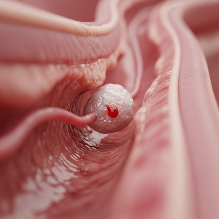
is a benign, non-cancerous tumour arising from the nerve sheath cells of peripheral nerves. It represents approximately 90% of all neurofibromas and typically occurs sporadically without association to genetic syndromes like neurofibromatosis type 1 (NF1). Unlike plexiform neurofibromas, which are pathognomonic for NF1, solitary forms are usually small, slow-growing, and asymptomatic flesh-coloured or pinkish bumps on or under the skin.These tumours develop centrally within the nerve, involving Schwann cells, fibroblasts, and perineural cells in a myxoid matrix. They are most common in young to middle-aged adults (ages 20–30), affecting males and females equally, and can appear anywhere on the body but favour extremities, trunk, and head/neck. Superficial cutaneous neurofibromas are pedunculated and extraneural, while deeper intraneural forms cause fusiform nerve expansion and may lead to neurological symptoms.
Rarely, solitary neurofibromas of deep-seated nerves (e.g., ulnar nerve) can signal underlying NF1 variants, necessitating genetic evaluation if café-au-lait spots or family history emerge. Malignant transformation to malignant peripheral nerve sheath tumour (MPNST) is exceedingly rare (<1%) in sporadic cases but higher in NF1.
Who gets solitary neurofibroma?
Solitary neurofibromas affect individuals across all ages but peak in the third decade of life. Approximately 95% arise sporadically in otherwise healthy people without NF1 or other genetic abnormalities.
- Demographics: Equal incidence in men and women; common in adults 20–50 years old.
- NF1 association: Absent in most cases (unlike multiple neurofibromas, a hallmark of NF1). However, deep solitary neurofibromas may prompt NF1 screening.
- Risk factors: Largely unknown; no strong environmental triggers identified. Sporadic mutations in nerve sheath cells implicated.
Children rarely present with solitary forms unless part of NF1 spectrum. Large or multiple lesions warrant genetic counselling.
What causes solitary neurofibroma?
The precise aetiology remains unclear, but solitary neurofibromas result from somatic mutations leading to uncontrolled proliferation of nerve sheath components. Unlike NF1-linked tumours (NF1 gene mutations on chromosome 17), sporadic cases lack germline defects.
- Pathogenesis: Involves Schwann cells producing excessive extracellular matrix (myxoid stroma); low cellularity with fusiform cells.
- Genetic factors: No familial pattern in 95% of cases. Rare NF1 gene variants (e.g., c.1722C>A) reported in atypical deep lesions.
- Non-genetic: Trauma or inflammation hypothesized but unproven.
Histologically, tumours are non-encapsulated, S100-positive (85%), and SOX10-reactive, distinguishing from schwannomas.
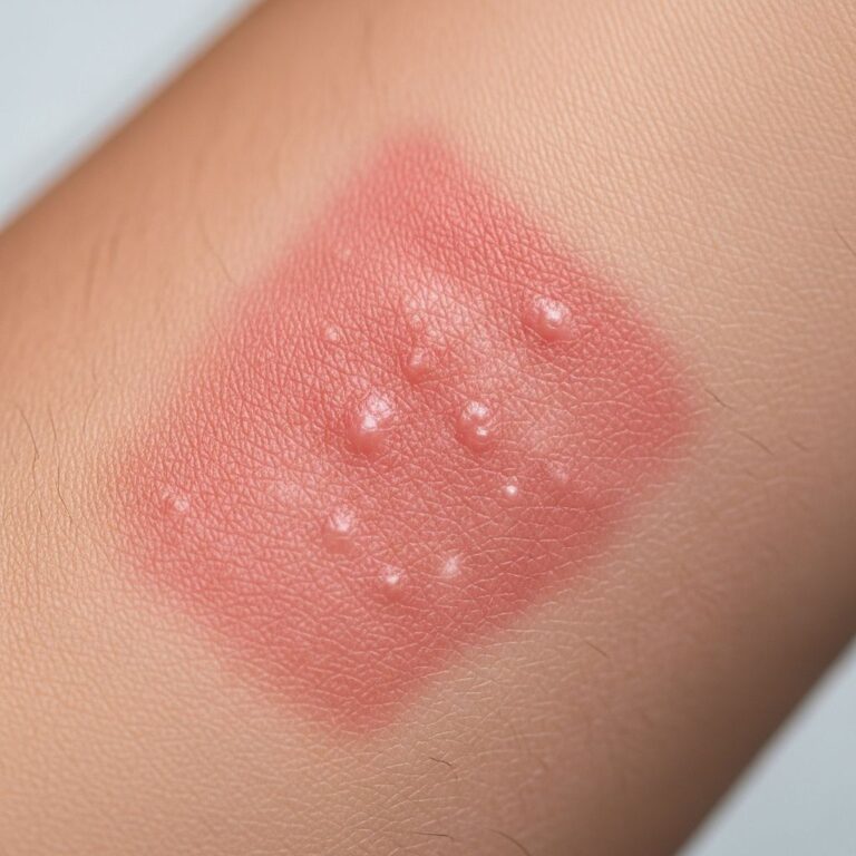
What are the clinical features of solitary neurofibroma?
Most solitary neurofibromas are asymptomatic, presenting as slow-growing, soft dermal or subcutaneous nodules (0.2–3 cm).
| Feature | Description |
|---|---|
| Appearance | Skin-coloured, pink, tan, or flesh-toned dome-shaped papule/nodule; soft, mobile, non-tender. |
| Symptoms | Usually none. Deep lesions: pain, paraesthesia, weakness if compressing nerves. |
| Surface | Smooth; rarely eroded/ ulcerated. Buttonhole sign: invagination on pressure. |
| Growth | Slow; may enlarge slightly over years but stabilizes. |
Superficial forms mimic dermatofibromas or lipomas; deep ones cause fusiform swelling with neurological deficits (e.g., numbness in thigh/ulnar distribution).
Diagnosis
Diagnosis relies on clinico-pathological correlation; excision biopsy is gold standard.
- Clinical: Characteristic soft nodule; rule out NF1 (café-au-lait spots, family history).
- Imaging: MRI preferred for deep lesions: T2-hyperintense “target sign” (central hypointensity), split-fat sign. Ultrasound: hypoechoic with posterior enhancement, vascularity. X-ray often normal.
- Histology: Fusiform cells in myxoid matrix; S100+ (less intense than schwannoma), collagenous zones; no Verocay bodies.
- Differentials: Schwannoma, leiomyoma, lipoma, fibroma, MPNST (rare).
Treatment
Observation suffices for asymptomatic lesions. Surgical excision for cosmetics, symptoms, or uncertainty.
- Conservative: Watchful waiting; low recurrence risk post-excision.
- Surgery: Complete excision with narrow margins; preserves nerve function in most. Postoperative: normal motion, no recurrence at 1-year.
- Other: No role for laser/radiofrequency routinely; emerging targeted therapies for NF1-linked (e.g., MEK inhibitors) not for solitary.
NF1 patients require lifelong monitoring for MPNST.
Complications
- Rare: Pain, neurological deficit (deep), cosmetic concern, malignant change (<1%).
- Surgery: Nerve injury, scarring.
- NF1 context: Multiple tumours, plexiform growth, MPNST risk.
Prevention
No known prevention; genetic counselling for NF1 families. Regular skin exams recommended.
Prognosis
Excellent; benign with minimal growth. Recurrence rare post-excision. Deep/NF1 cases need surveillance.
Frequently Asked Questions
Q: Is solitary neurofibroma cancerous?
A: No, it is benign. Malignant transformation is extremely rare in sporadic cases.
Q: Should I have it removed?
A: Only if symptomatic, growing, or for cosmetics. Observation is safe.
Q: Can it be linked to neurofibromatosis?
A: Rarely; screen for NF1 if deep, multiple, or with café-au-lait spots.
Q: What does it look like on MRI?
A: Target sign on T2, split-fat sign; hypoechoic on ultrasound.
Q: Does it hurt?
A: Usually not; deep ones may cause pain/numbness.
References
- Atypically Located Solitary Neurofibroma of the Ulnar Nerve — PMC/NCBI. 2022. https://pmc.ncbi.nlm.nih.gov/articles/PMC9492796/
- Neurofibroma – Symptoms and causes — Mayo Clinic. 2023-10-12. https://www.mayoclinic.org/diseases-conditions/neurofibroma/symptoms-causes/syc-20573693
- Neurofibroma — Cleveland Clinic. 2023. https://my.clevelandclinic.org/health/diseases/22535-neurofibroma
- Neurofibroma Solitary — Buffalo Medical Group. 2017. https://www.buffalomedicalgroup.com/wp-content/uploads/2017/08/Neurofibroma-Solitary.pdf
- Solitary Neurofibroma in Lower Extremity — Brieflands. 2015. https://brieflands.com/journals/ijcm/articles/9342
Similar Articles


Read full bio of medha deb