Specific Viral Exanthems: Diagnostic Guide For Pediatricians
Comprehensive guide to recognising and managing classic childhood viral rashes with distinctive morphologies.

An
exanthem
is a widespread rash, usually of viral origin, accompanied by systemic symptoms such as fever, headache, and malaise. Specific viral exanthems are childhood eruptions with distinctive morphologies that allow clinical diagnosis. This article reviews their key features, aiding dermatologists and paediatricians in accurate identification.Introduction to Viral Exanthems
Viral exanthems represent a significant proportion of paediatric consultations, often presenting with fever followed by characteristic rashes. These rashes arise from immune responses to viral antigens rather than direct cytopathic effects. Common viruses include enteroviruses, parvovirus B19, and herpesviruses. Diagnosis relies on rash morphology, distribution, and associated symptoms, with historical vaccination reducing incidence of classic exanthems like measles.
Key diagnostic principles include age of onset (typically under 10 years), prodromal fever, and rash evolution. Differentials encompass bacterial infections, drug eruptions, and autoimmune conditions. Management is supportive, focusing on hydration, antipyretics, and itch relief.
Measles
Measles, caused by the measles virus (Morbillivirus), remains a global concern despite vaccination. Prodrome includes high fever, cough, coryza, conjunctivitis (the ‘3 Cs’), and Koplik spots—pathognomonic 1–2 mm white lesions on buccal mucosa opposite molars, appearing 1–2 days before rash.
The
exanthem
starts as erythematous macules behind ears and on face, spreading cephalocaudally to trunk and extremities over 3–4 days. Lesions coalesce into blotches, blanching on pressure. Rash fades in 5–7 days with desquamation, often leaving brownish pigmentation.Complications include pneumonia, encephalitis (1/1000 cases), and subacute sclerosing panencephalitis. Diagnosis is clinical; PCR confirms virus. Notify public health authorities. Differentials: Kawasaki disease, scarlet fever.
Rubella
Rubella (German measles), due to rubella virus, presents milder than measles. Prodrome: low-grade fever, sore throat, retroauricular lymphadenopathy (posterior triangle hallmark). Exanthem comprises discrete pink macules/papules starting on face, spreading to trunk/limbs in 24 hours. Fades in 3 days without desquamation.
Forchheimer spots (palatal petechiae) occur in 20–50%. In pregnancy, congenital rubella syndrome risks deafness, cataracts, heart defects. Serology or PCR diagnoses. Differentials: scarlet fever, enteroviral exanthems. Vaccination prevents most cases.
Roseola Infantum (Exanthem Subitum)
Human herpesvirus 6 (HHV-6) or HHV-7 causes roseola, affecting infants 6–24 months. High fever (39–40°C) lasts 3–5 days, with febrile seizures in 10–15%. As fever resolves, pink-red blanching macules/papules erupt on trunk, spreading to neck, face, extremities. Resolves in 1–2 days.
Mild upper respiratory symptoms, irritability common. Diagnosis clinical; HHV-6 PCR if atypical. Differentials: measles, Kawasaki. Supportive care; antipyretics (avoid aspirin).
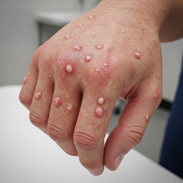
Hand, Foot, and Mouth Disease
Primarily Coxsackie A16 or Enterovirus 71. Prodrome: fever, malaise, sore throat. Oral lesions: 2–5 mm grey-white vesicles/ulcers on anterior tongue, buccal mucosa. Skin: oval grey vesicles with red halo on palms, soles, buttocks; may generalize in outbreaks.
Evolves to crusts in 7–10 days; post-infectious onychomadesis (nail shedding) in 4–8 weeks. Severe EV71 cases: neurological complications. PCR from vesicles/stool confirms. Differentials: eczema herpeticum, bullous impetigo.
Pityriasis Rosea
Possibly human herpesvirus 6/7-associated. ‘Herald patch’: 2–5 cm oval salmon-pink plaque with collarette scale on trunk/abdomen. 1–2 weeks later, smaller plaques (‘Christmas tree’ pattern) on trunk. Christmas tree distribution on back with peripheral trailing scale.
Mild itch/pruritus; resolves 6–8 weeks. Oral lesions rare. Diagnosis clinical; biopsy if atypical. Differentials: guttate psoriasis, tinea corporis. Topical steroids for itch.
Fifth Disease (Erythema Infectiosum)
Parvovirus B19 causes ‘slapped cheek’ appearance: bright red confluent erythema on cheeks sparing nasolabial folds/circumoral. 1–4 days later, lacy reticular erythema on proximal extremities, trunk. Rash recurs with heat/sun/exercise for weeks.
Arthralgias in adults; aplastic crisis in sickle cell. Serology diagnoses. Avoid in pregnancy (hydrops fetalis risk). Differentials: drug eruption.
Papular Acrodermatitis (Gianotti-Crosti Syndrome)
Clinical Features
Symmetrical monomorphic papules (1–5 mm) on cheeks, buttocks, extensor limbs; trunk spared. Triggered by hepatitis B, EBV, Coxsackie. Lasts 2–10 weeks; lymphadenopathy, rarely liver dysfunction.
Investigations
Hep B serology if unvaccinated; LFTs. Biopsy shows spongiotic dermatitis.
Differential Diagnosis
- Insect bite reactions
- Scabies
- Papular urticaria
- Lichen planus
Varicella (Chickenpox)
Primary varicella-zoster virus (VZV) infection. Prodrome mild fever. Centripetal pruritic vesicles: ‘dew drop on rose petal’—papule to vesicle to pustule to crust. Successive crops over 5–7 days; 200–500 lesions.
Complications: bacterial superinfection, pneumonia (adults/smokers). PCR from swab diagnoses. Aciclovir for severe/immunocompromised. Isolation until crusted.
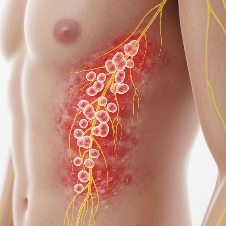
Herpes Zoster (Shingles)
VZV reactivation. Prodrome: dermatomal pain, paraesthesia 3–5 days. Grouped vesicles on erythematous base in dermatome (thoracic T5-L2 common). Crusts in 7–10 days; post-herpetic neuralgia risk in elderly.
PCR confirms. Antivirals (aciclovir/valaciclovir) within 72h reduce duration. Differentials: herpes simplex, contact dermatitis.
Other Viral Exanthems
Enteroviral Exanthems
Echovirus/Coxsackie: morbilliform, petechial, urticarial rashes. Summer epidemics.
Non-Specific Childhood Exanthem
Most common; post-infectious maculopapular rash.
Drug Eruptions Mimicking Exanthems
Amoxicillin common mimic.
Diagnostic Approach
History: Fever duration, contacts, vaccination, exposures.
Examination: Rash distribution/morphology, lymphadenopathy, mucosal lesions.
Investigations: Selective PCR/serology; FBC, CRP if systemic.
| Exanthem | Incubation | Rash Distribution | Key Feature |
|---|---|---|---|
| Measles | 10–14 days | Face to trunk | Koplik spots |
| Rubella | 14–21 days | Face/trunk | Retroauricular nodes |
| Roseola | 5–15 days | Trunk | Post-febrile |
| HFMD | 3–6 days | Hands/feet/mouth | Vesicles on palms |
| Fifth Disease | 13–18 days | Cheeks/extremities | Slapped cheek |
Frequently Asked Questions (FAQs)
What causes viral exanthems?
Primarily viruses like measles, rubella, parvovirus B19, enteroviruses, and herpesviruses trigger immune-mediated rashes.
How are viral exanthems diagnosed?
Clinical features suffice for classics; PCR/serology for confirmation.
Is treatment needed for viral rashes?
Supportive: paracetamol, hydration, calamine. Antivirals for severe varicella/zoster.
Can viral exanthems be prevented?
Yes, via MMR vaccine for measles/rubella/varicella.
When to seek urgent care?
High fever >5 days, breathing difficulty, neurological signs, or immunocompromise.
References
- Clinical features of viral exanthems — Royal Australian College of General Practitioners. 2021-04-01. https://www1.racgp.org.au/ajgp/2021/april/clinical-features-of-viral-exanthems
- Viral Exanthems Rashes — Children’s National Hospital. 2024. https://www.childrensnational.org/get-care/health-library/viral-exanthems-rashes
- Viral Exanthem Rash: Symptoms, Causes & Treatment — Cleveland Clinic. 2023-10-12. https://my.clevelandclinic.org/health/diseases/22510-viral-exanthem-rash
Similar Articles


Read full bio of Sneha Tete