Spider Telangiectasis: Causes, Diagnosis, and Treatment
Comprehensive guide to spider telangiectasis: Understanding dilated blood vessels, clinical features, and treatment options.

Spider Telangiectasis: A Comprehensive Overview
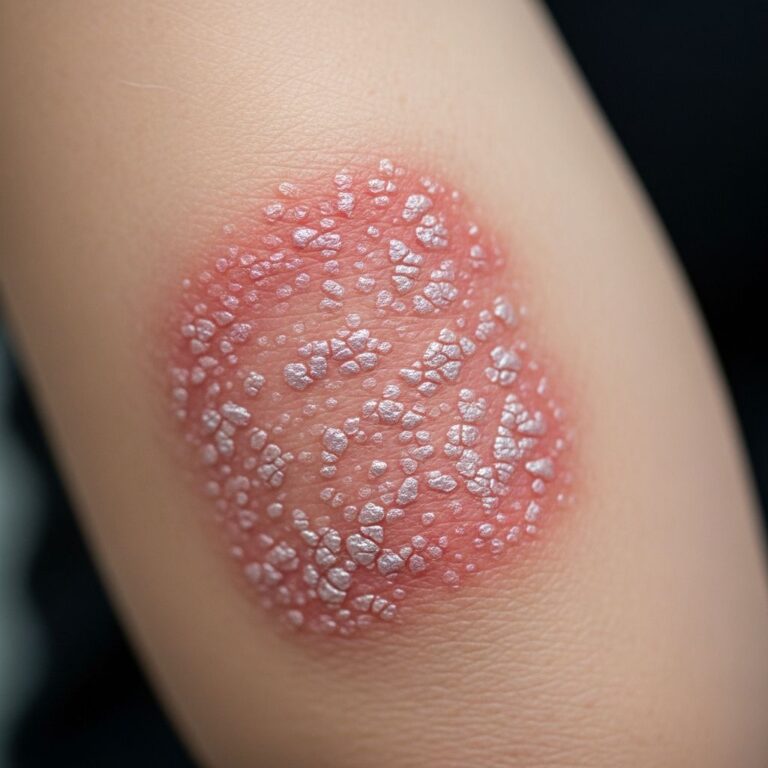
A spider telangiectasis is a common vascular lesion composed of dilated blood vessels that presents with a distinctive spider-like appearance. The condition is characterized by a central red papule (representing the ‘body’ of the spider) from which fine red lines extend radially outward, resembling spider legs. Alternative names for this condition include spider angioma, spider naevus, and naevus araneus. The lesion’s name derives from either its central red papule with radiating extensions or the spider-like network pattern formed by the red lines themselves.
Epidemiology and Prevalence
Spider telangiectasis is a remarkably common condition affecting a significant portion of the population. A solitary spider telangiectasis affects approximately 10–15% of both children and adults. While individuals of any race can develop these lesions, they are more readily visible in individuals with fair skin compared to those with darker skin tones.
Multiple spider telangiectases, however, are associated with specific medical and physiological conditions. These include:
- Pregnancy
- Use of combined oral contraceptive pills
- Liver disease, particularly cirrhosis resulting from alcohol abuse
- Thyrotoxicosis
The presence of multiple spider angiomas is highly specific for chronic liver disease, with a specificity of 95%. This makes their presence an important clinical indicator when evaluating patients with suspected hepatic dysfunction.
Pathophysiology and Mechanisms
Understanding how spider telangiectasis develops requires knowledge of the underlying vascular changes that occur. Spider telangiectasis is classified as an acquired vascular malformation that develops due to the failure of a tiny muscle (the sphincteric muscle) that normally restricts the size of an arteriole. This muscular failure leads to significant changes in blood flow dynamics.
When the sphincteric muscle fails, increased pulsating flow occurs through the central vessel (the papule), resulting in the dilatation of distal vessels (the red lines extending outward). The exact mechanism triggering this initial muscular failure has not been completely elucidated, but several important hypotheses have been proposed.
Hormonal factors play a prominent role in spider telangiectasis development. The condition may arise spontaneously or be induced by circulating estrogen, which is elevated in several conditions:
- Pregnancy
- Combined oral contraceptive use
- Liver disease
Additional mechanisms implicated in pathogenesis include direct vasodilatory effects of alcohol, substance P, hyperestrogenism, and inadequate hepatic metabolism of steroid hormones. Recent research has identified elevated serum vascular growth factors, particularly vascular endothelial growth factor (VEGF) and basic fibroblast growth factor (bFGF), in patients with liver cirrhosis, suggesting that angiogenesis may contribute to spider nevi formation.
Clinical Features and Presentation
Spider telangiectases present with characteristic clinical features that aid in their recognition and diagnosis. The typical appearance consists of a central dilated arteriole from which numerous thin-walled capillary branches radiate, creating the spider-like configuration.
Location patterns are an important clinical consideration. Spider telangiectases are frequently located on:
- The face
- The neck
- The upper chest
- The hands and arms
- Other body sites
The predominance on the face, neck, and upper chest has been postulated to relate to the distribution of the superior vena cava, a large vein draining the heart.
Morphological variations are commonly observed. Spider telangiectases vary considerably in size and number, with lesions tending to be larger and more numerous in individuals with severe liver disease. In such cases, other cutaneous signs of liver disease may be present, including palmar erythema, leukonychia, and jaundice. The central dilated arteriole may sometimes be present without radial capillaries, and the capillaries themselves may vary in diameter, length, and number, occasionally appearing star-shaped rather than spider-like.
Symptoms and complications are minimal in most cases. The lesions may briefly bleed on trauma but otherwise do not cause any symptoms or complications in the vast majority of patients.
Diagnosis
Spider telangiectasis diagnosis is primarily based on clinical appearance. The distinctive spider-like morphology is usually sufficient for diagnosis without requiring additional testing in straightforward cases.
A simple clinical maneuver can confirm the diagnosis and demonstrate the vascular nature of the lesion. Compression of the central arteriole results in the rapid disappearance of the radial capillaries, which quickly refill when compression is relieved. This blanching and refilling phenomenon is best visualized through a transparent object, such as a glass slide or the lens of a contact dermatoscope. This test effectively confirms the vascular origin of the lesion and distinguishes it from other skin lesions with similar appearance.
Differential Diagnoses
While spider telangiectasis has a characteristic appearance, several other skin conditions may present similarly and should be considered in the differential diagnosis:
- Hereditary hemorrhagic telangiectasia (Osler-Weber-Rendu syndrome)
- Rosacea with telangiectasia
- Varicose veins
- Port-wine stains (capillary malformations)
- Other vascular malformations
Distinguishing features, particularly the presence of a central arteriole with radiating capillaries visible on dermatoscopy, help differentiate spider telangiectasis from these other conditions.
Treatment Options
While spider telangiectasis is generally benign and asymptomatic, many patients seek treatment for cosmetic reasons. Several treatment modalities are available:
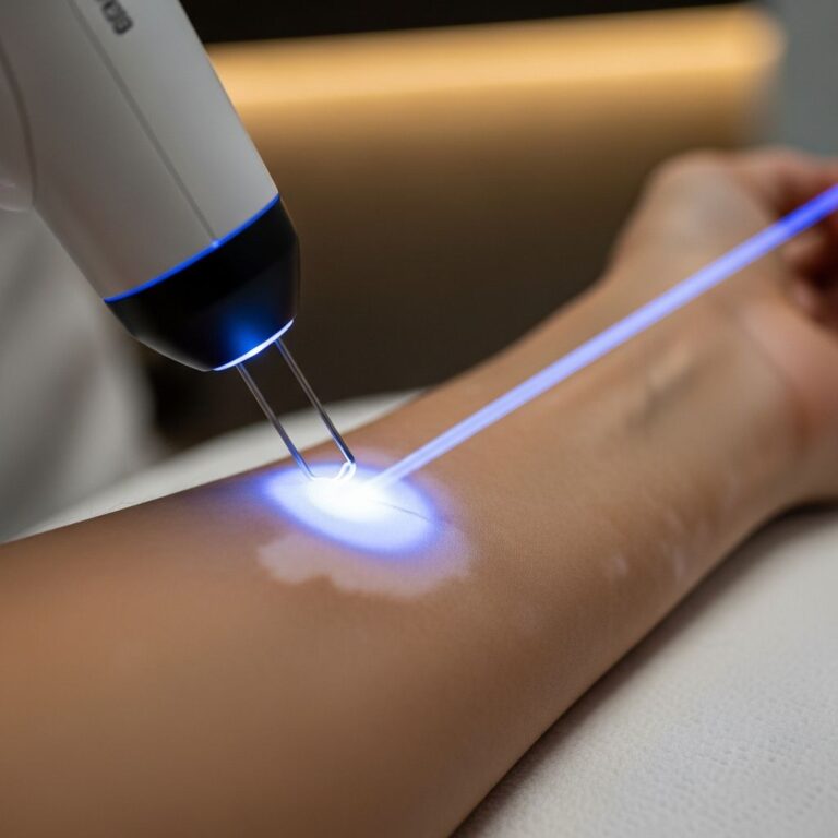
- Laser therapy – Utilizes targeted light energy to collapse dilated vessels
- Electrocautery – Uses electrical current to treat the central arteriole
- Sclerotherapy – Involves injection of sclerosing agents to close affected vessels
- Cryotherapy – Uses freezing to destroy the vascular lesion
The choice of treatment depends on factors including lesion size, location, patient skin type, and patient preference. Many lesions can be effectively treated with a single procedure, though some may require multiple sessions for optimal results.
Associated Medical Conditions
The presence of spider telangiectasis, particularly when multiple lesions are present, should prompt evaluation for underlying systemic disease. Multiple spider angiomas are highly suggestive of:
- Cirrhosis – Most commonly associated with chronic liver disease
- Rheumatoid arthritis
- Thyrotoxicosis
In patients with multiple spider telangiectases without an obvious cause (such as pregnancy or oral contraceptive use), thorough clinical evaluation for liver disease and other systemic conditions is warranted.
Risk Factors and Predisposing Conditions
Several factors increase the likelihood of developing spider telangiectasis:
- Genetic predisposition – Family history of vascular conditions increases susceptibility
- Pregnancy – Increased red blood cells and elevated estrogen/progesterone
- Hormonal medications – Combined oral contraceptives and hormone replacement therapy
- Chronic liver disease – Impaired metabolism of steroid hormones
- Chronic systemic or topical corticosteroid use
- Sun damage – Particularly in fair-skinned individuals
- Trauma or injury to blood vessels
Outcome and Natural History
The natural history of spider telangiectasis varies depending on the underlying etiology. Solitary lesions in otherwise healthy individuals may remain stable or may gradually enlarge over time. Pregnancy-related lesions often regress partially or completely in the postpartum period as hormone levels normalize. In contrast, lesions associated with chronic liver disease tend to be progressive and may increase in number and size as the underlying hepatic dysfunction worsens.
Spontaneous resolution of spider telangiectasis is relatively uncommon, particularly in solitary lesions in healthy individuals or in those with underlying systemic disease. Therefore, most patients seeking resolution of these lesions require active treatment.
Frequently Asked Questions
Q: Is spider telangiectasis dangerous?
A: Spider telangiectasis is generally benign and does not pose a health threat. However, the presence of multiple lesions may indicate underlying systemic disease such as liver disease, requiring further medical evaluation.
Q: Can spider telangiectasis be prevented?
A: While genetic predisposition cannot be changed, certain risk factors can be minimized. These include limiting sun exposure, avoiding unnecessary topical steroid use, managing underlying medical conditions, and discussing hormonal contraceptive alternatives with a healthcare provider if concerned about vascular lesions.
Q: Will spider telangiectasis treated with laser return?
A: Most successfully treated spider telangiectases do not recur at the same location. However, new spider telangiectases may develop elsewhere if the underlying risk factors (such as pregnancy or continued oral contraceptive use) persist.
Q: Are spider telangiectases more common in certain populations?
A: While spider telangiectases can occur in individuals of any race, they are more easily visible in fair-skinned individuals compared to those with darker skin tones. The actual prevalence may be similar across populations, but visibility differs.
Q: What should I do if I suddenly develop multiple spider telangiectases?
A: The sudden onset of multiple spider telangiectases warrants medical evaluation, particularly to assess for liver disease, particularly if you are not pregnant or taking oral contraceptives. Consultation with a dermatologist or primary care physician is recommended.
References
- Spider telangiectasis — DermNet New Zealand. Accessed January 2026. https://dermnetnz.org/topics/spider-telangiectasis
- What Are Telangiectasias? Causes and Treatment for Spider Veins — GoodRx Health. Accessed January 2026. https://www.goodrx.com/health-topic/dermatology/telangiectasia-spider-veins-causes-treatment
- Spider Angioma — StatPearls, National Center for Biotechnology Information (NCBI). Accessed January 2026. https://www.ncbi.nlm.nih.gov/books/NBK507818/
- Spider veins: Treatment, causes and everything else you need to know — Mercy One. Accessed January 2026. https://www.mercyone.org/blog-articles/spider-veins-treatment-causes-and-everything-else-you-need-know
- Telangiectasia: Causes, Diagnosis, Prevention & Treatment — Hospital for Special Surgery (HSS). Accessed January 2026. https://www.hss.edu/health-library/conditions-and-treatments/telangiectasia-and-autoimmune-disease
Similar Articles


Read full bio of Sneha Tete