Spitzoid Melanoma: Clinical Features, Diagnosis, and Treatment
Understanding spitzoid melanoma: A deceptive skin cancer that mimics benign moles.

What is Spitzoid Melanoma?
Spitzoid melanoma is a malignant melanoma that presents a significant diagnostic challenge due to its histological similarity to a benign skin lesion known as Spitz naevus. This rare form of cutaneous melanoma accounts for less than 2% of all melanomas, yet it represents one of the most common types of melanomas in children. The condition occurs when irregular mutations in DNA cause melanocytes to become cancerous and grow uncontrollably, most commonly triggered by extended exposure to ultraviolet (UV) rays from the sun or tanning beds.
The primary challenge in managing spitzoid melanoma lies in its deceptive appearance and microscopic features that closely resemble benign Spitz naevi. Unlike other melanoma types that typically present with asymmetry, irregular borders, and color variation, spitzoid melanoma is often round in shape and uniform in colour, failing to follow the commonly used ABCDE criteria for melanoma detection. This disguising presentation can delay diagnosis and complicate clinical management, making awareness and vigilance essential for healthcare providers.
Clinical Presentation and Features
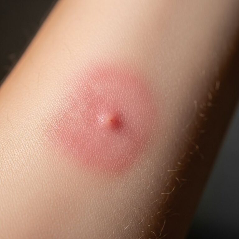
Spitzoid melanoma presents as a changing and enlarging papule or nodule that warrants clinical attention. The lesion can manifest in various colors, appearing as either amelanotic (nonpigmented, red or pink) or pigmented (brown, black, or blue). In advanced stages, spitzoid melanoma may become crusted and ulcerated, signaling disease progression.
The anatomical distribution of spitzoid melanoma shows a predilection for the head and extremities, though it can arise on any body location and in any ethnic group. Most commonly, these lesions are found on the arms, legs, or face. Notably, spitzoid melanoma can arise de novo as a new lesion, or it may develop within an existing Spitz naevus, adding another layer of diagnostic complexity.
Key distinguishing clinical features include:
- Round or uniform shape, unlike the asymmetry typical of other melanomas
- Uniform or relatively uniform coloration
- Rapid growth patterns, often enlarging noticeably over weeks to months
- Small initial size, frequently less than 6mm in diameter initially
- Dome-shaped appearance with well-defined borders
- May be surrounded by inflammatory changes or erythema
Histopathological Features
The histological diagnosis of spitzoid melanoma requires careful microscopic examination by experienced pathologists, as the features overlap considerably with benign Spitz naevi. Compared to Spitz naevus, spitzoid melanoma demonstrates several distinguishing characteristics that aid in differentiation.
Cytologically, spitzoid melanomas display high nuclear to cytoplasmic ratios with dusty cytoplasmic melanisation along with large eosinophilic nucleoli. The histologic features that characterize these lesions include:
- Hyperkeratosis and patchy parakeratosis in the epidermis
- Presence of large acidophilic (eosinophilic) cells
- Mononuclear or multinuclear giant cells
- Occasional mitotic figures, which may be increased compared to benign variants
- Pigment located predominantly in the superficial part of the lesion
- Inflammatory changes and edema in the dermis and epidermis
- Capillary dilation in the papillary dermis
- Junctional nests with variable architectural patterns
- Atypical melanocytic proliferation with increased cellularity
These histological findings must be interpreted within the clinical context, as some atypical Spitz tumors cannot be definitively classified as benign or malignant. In such cases, the lesion is designated as a Spitzoid Tumor of Uncertain Malignant Potential (STUMP), reflecting diagnostic uncertainty and the need for close clinical follow-up.
Diagnostic Approach
Diagnosis of spitzoid melanoma is established through skin biopsy of an enlarging nodule. When a clinician identifies a suspicious spitzoid lesion, complete excisional biopsy is typically recommended to obtain adequate tissue for comprehensive histological evaluation. The biopsy specimen should be carefully examined under the microscope by a dermatopathologist experienced in evaluating melanocytic lesions.
A healthcare provider uses skin biopsy to obtain diagnostic material. During the biopsy procedure, the entire skin growth is removed or a representative sample is obtained, and the specimen is sent to a laboratory where a pathologist evaluates it for signs of skin cancer. The pathological examination must assess not only the presence of malignancy but also determine the Breslow thickness, mitotic rate, and presence or absence of ulceration, as these factors influence prognosis and treatment decisions.
Additional imaging and staging studies may be recommended if spitzoid melanoma is confirmed:
- Sentinel lymph node biopsy (SLNB) in selected cases to determine regional lymph node involvement
- Imaging studies (CT, PET, or MRI) for thicker tumours or those with concerning features
- Clinical assessment and documentation of all suspicious lesions
- Photography for baseline comparison and follow-up monitoring
The role of sentinel lymph node biopsy in atypical spitzoid melanocytic lesions remains controversial, and the reliability of associated prognostic information is unclear. However, patients with spitzoid melanoma and positive sentinel lymph node biopsy have been shown to have a more indolent disease course than those with positive sentinel lymph node biopsy and non-spitzoid melanomas, suggesting biological differences in this melanoma subset.
Differential Diagnosis
The primary diagnostic challenge in spitzoid melanoma involves distinguishing it from benign Spitz naevus. Spitz naevi are benign melanocytic proliferations that typically present as rapidly growing papules in individuals under 35 years of age. Classic and pigmented Spitz nevi are considered harmless, though some eventually disappear, usually leaving only a temporary area of discoloration.
However, some atypical Spitz tumors have been linked to melanoma, though researchers are not entirely sure how these conditions relate. Very few atypical Spitz tumors progress to melanoma and lead to serious health risks, especially when diagnosed and treated early. The distinction between benign Spitz naevus, atypical Spitz nevus, STUMP, and spitzoid melanoma requires experienced pathological interpretation and may necessitate additional molecular studies or immunohistochemical analysis to guide clinical management.
Treatment Approach
Spitzoid melanoma should be completely excised with a margin of normal tissue surrounding the lesion. The recommended clinical margins depend on the measured thickness of the tumour, following standard melanoma surgical principles. Wide local excision with appropriate margins is the mainstay of treatment.
The treatment approach is generally based on the following principles:
- Complete surgical excision with adequate margins (typically 1-2 cm for thin melanomas, 2 cm for thicker lesions)
- Histopathological examination of the entire specimen to confirm complete removal and assess for adverse prognostic features
- Sentinel lymph node biopsy consideration in intermediate to thick lesions
- Adjuvant therapy evaluation based on stage and risk factors in appropriate candidates
- Close clinical and dermatological follow-up with regular surveillance
Unlike benign Spitz nevi, which may be monitored clinically if classic features are present, spitzoid melanoma requires definitive surgical treatment. When spitzoid melanoma is confirmed histologically, prompt wide local excision with appropriate margins is essential to prevent local recurrence and reduce the risk of regional or distant metastasis.
In thicker tumours, sentinel lymph node biopsy may be recommended to determine whether the melanoma has spread to regional lymph nodes. This information is crucial for staging, prognostication, and guiding decisions regarding adjuvant therapy. However, as mentioned previously, the role and prognostic significance of SLNB in atypical spitzoid lesions remains an area of ongoing clinical investigation and debate.
Prognosis and Outlook
Spitzoid melanoma is usually less aggressive than other types of melanomas, even though the moles may grow fairly rapidly. This relatively indolent biological behavior, particularly compared to other melanoma subtypes, represents an important prognostic distinction. However, the sooner any melanoma is detected, the better the chances of successful treatment.
The overall prognosis depends on several factors:
- Breslow Thickness: Thinner lesions (less than 1mm) generally have significantly better outcomes
- Mitotic Rate: Higher mitotic activity may indicate more aggressive behaviour
- Ulceration: Presence of ulceration is associated with worse prognosis
- Stage at Diagnosis: Early-stage lesions without nodal or distant metastases have excellent prognosis
- Sentinel Lymph Node Status: Patients with positive SLNB require closer surveillance and may benefit from adjuvant therapy
- Age: Young patients, particularly children, tend to have more favorable outcomes
Early detection and treatment significantly improve outcomes. Most patients with stage I spitzoid melanoma (localized disease without nodal involvement) have excellent long-term survival rates when appropriately treated with complete surgical excision. Regional and distant recurrence rates are generally lower in spitzoid melanoma compared to other melanoma types, though individual cases may vary.
Risk Factors and Prevention
While the most common cause of spitzoid melanoma involves UV mutations in DNA resulting from extended exposure to ultraviolet rays from the sun or tanning beds, certain individuals carry increased susceptibility.
In rare cases, individuals may inherit a gene mutation that increases melanoma risk. For example, people with xeroderma pigmentosum (XP), an inherited genetic condition, have a mutation that impairs their bodies’ ability to repair DNA damage after UV exposure, significantly increasing melanoma risk.
Prevention strategies include:
- Limiting sun exposure, particularly during peak hours (10 AM to 4 PM)
- Consistent use of broad-spectrum sunscreen (SPF 30 or higher)
- Wearing protective clothing, hats, and sunglasses
- Avoiding tanning beds and artificial UV sources
- Regular self-examination of skin for changing moles or new lesions
- Professional skin examinations by dermatologists, particularly for high-risk individuals
- Genetic counselling for individuals with family history of melanoma
Special Considerations in Pediatric Cases
Spitzoid melanoma is one of the most common types of melanomas in children, despite melanoma being generally rare in this age group. The presentation of rapidly growing pigmented lesions in children warrants prompt evaluation by experienced dermatologists. Spitz naevi are also common in children, and distinguishing benign lesions from spitzoid melanoma in this population requires particular expertise.
Management principles in children remain similar to adults, emphasizing complete surgical excision of suspicious lesions. However, the biological behavior and prognosis may differ, with many pediatric cases demonstrating indolent courses even when diagnosed as spitzoid melanoma. Close long-term follow-up is essential in all pediatric cases.
Frequently Asked Questions
Q: How does spitzoid melanoma differ from regular melanoma?
A: Spitzoid melanoma differs from typical melanoma in its appearance and behaviour. It is usually round and evenly colored, not asymmetrical or irregularly pigmented like classic melanomas. Additionally, spitzoid melanoma tends to be less aggressive and has better long-term prognosis when detected and treated early, particularly in pediatric cases.
Q: Can spitzoid melanoma arise from a benign Spitz naevus?
A: Yes, spitzoid melanoma can develop within an existing Spitz naevus, though it can also arise de novo as a new lesion. This possibility underscores the importance of monitoring changing or enlarging spitzoid lesions and obtaining biopsy when clinical features suggest malignant transformation.
Q: What is the role of sentinel lymph node biopsy in spitzoid melanoma?
A: The role of sentinel lymph node biopsy in spitzoid melanoma is somewhat controversial, and the reliability of prognostic information from SLNB in atypical spitzoid lesions remains unclear. However, it may be recommended for thicker tumours to determine if cancer has spread to regional lymph nodes, which helps guide treatment decisions and adjuvant therapy options.
Q: Why is spitzoid melanoma difficult to diagnose?
A: Spitzoid melanoma is difficult to diagnose because it closely resembles benign Spitz naevus histologically and does not follow the typical ABCDE criteria for melanoma. Its round shape, uniform color, and benign-appearing microscopic features can be misleading even to experienced pathologists, sometimes leading to diagnostic uncertainty requiring molecular testing or close clinical follow-up.
Q: What is the prognosis for patients with spitzoid melanoma?
A: Spitzoid melanoma generally has a better prognosis than other melanoma types, particularly when detected and treated early. Early-stage disease with complete surgical excision and appropriate margins offers excellent long-term survival. However, prognosis depends on factors including lesion thickness, mitotic rate, ulceration, and presence of nodal involvement.
Q: What age group is most commonly affected by spitzoid melanoma?
A: While all age groups can develop spitzoid melanoma, it is one of the most common types of melanomas in children. However, it can occur at any age. Any individual noticing a rapidly growing pigmented lesion should seek medical evaluation regardless of age.
References
- Spitzoid Melanoma — DermNet NZ. 2024. https://dermnetnz.org/topics/spitzoid-melanoma
- Spitzoid Melanoma: Symptoms, Diagnosis, Staging & Treatment — Cleveland Clinic. 2024. https://my.clevelandclinic.org/health/diseases/24153-spitzoid-melanoma
- Types of Melanoma and What They Mean — Healthgrades. 2024. https://resources.healthgrades.com/right-care/melanoma/types-of-melanoma-and-what-they-mean
- Types of Melanoma with Pictures — Healthline. 2024. https://www.healthline.com/health/skin-cancer/types-of-melanoma
- Spitz Nevus: Pictures, Diagnosis, and Treatment — Medical News Today. 2024. https://www.medicalnewstoday.com/articles/320805
- Sophie Spitz: A Woman Ahead of Her Time — PubMed Central. 2019. https://pmc.ncbi.nlm.nih.gov/articles/PMC6637087/
- Genetics of Skin Cancer — National Cancer Institute. 2024. https://www.cancer.gov/types/skin/hp/skin-genetics-pdq
Similar Articles


Read full bio of Sneha Tete