Squamous Cell Carcinoma of the Ear Images
Comprehensive visual guide to squamous cell carcinoma on the ear, covering clinical images, symptoms, diagnosis, and pathology for dermatologists and patients.

Squamous cell carcinoma (SCC) is the most common malignant tumour of the ear. This chapter incorporates the most salient clinical features and images of squamous cell carcinoma of the ear, highlighting pinna, external auditory canal, and middle ear involvement.
What is squamous cell carcinoma of the ear?
Squamous cell carcinoma of the ear arises from the squamous epithelium lining the pinna, external auditory canal (EAC), or middle ear. It represents a significant proportion of non-melanoma skin cancers on sun-exposed ear sites and rare primary malignancies within the temporal bone. Chronic inflammation, such as long-standing otitis media or externa, predisposes to malignant transformation, often presenting at advanced stages due to delayed diagnosis.
On the pinna, SCC typically manifests as an ulcerated nodule or plaque on the helix or antihelix, frequently in fair-skinned individuals with cumulative UV exposure. In the EAC, it erodes cartilage and bone, invading the middle ear, facial nerve, parotid gland, or temporomandibular joint (TMJ), with potential intracranial extension. Middle ear SCC is exceedingly rare but aggressive, spreading via mastoid air cells or internal auditory canal.
Who gets squamous cell carcinoma of the ear?
SCC of the ear predominantly affects older adults, particularly those over 60, with a male predominance due to occupational sun exposure (e.g., farmers, sailors) and lighter skin types (Fitzpatrick I–III). Risk factors include:
- Chronic sun exposure and UV radiation
- Immunosuppression (e.g., organ transplant recipients)
- Chronic otitis externa/media or prior radiation
- Human papillomavirus (HPV) infection, especially high-risk types
- Arsenic exposure or chemical carcinogens
- Previous skin cancers or actinic keratoses
In the EAC and temporal bone, patients often have a history of chronic ear infections, with symptoms misattributed to refractory otitis for months before biopsy confirmation.
What causes squamous cell carcinoma of the ear?
The pathogenesis involves genetic mutations in squamous keratinocytes triggered by environmental and host factors. Ultraviolet B radiation induces p53 mutations, leading to uncontrolled proliferation. In chronically inflamed ears, squamous metaplasia progresses to dysplasia and invasive carcinoma.
Key causative factors:
- UV radiation: Primary driver for pinna SCC, with fair skin and outdoor occupations increasing risk.
- Chronic inflammation: Otitis media/externa promotes metaplasia; EAC SCC often overlays granulation tissue.
- HPV: High-risk subtypes (16, 18) implicated in mucosal SCC of EAC/middle ear.
- Immunosuppression: Increases incidence 65–250-fold in transplant patients.
What are the clinical features of squamous cell carcinoma of the ear?
Clinical presentation varies by site. Pinna lesions are often early-detected due to visibility, while EAC/middle ear tumours present late with destructive symptoms.
Pinna SCC
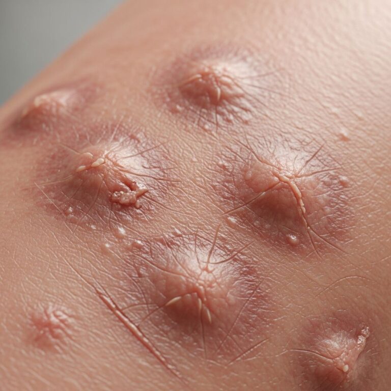
Commonly appears as a firm, hyperkeratotic nodule, ulcer, or plaque on the helix, scaphoid fossa, or antihelix. Lesions are pink-red, scaly, and may bleed or crust. Advanced cases show rolled borders, central ulceration, and fixation to cartilage.
- Early: Scaly patch or wart-like growth
- Advanced: Ulcerated tumour >2 cm, perichondrial invasion
External Auditory Canal (EAC) SCC
Symptoms mimic chronic otitis: persistent otorrhea (serous, purulent, blood-stained), otalgia, conductive hearing loss, and granulation tissue in the canal. Facial palsy indicates facial nerve involvement; trismus suggests TMJ invasion.
- Discharge unresponsive to antibiotics
- Temporal bone erosion on exam
- Mass protruding from canal
Middle Ear/Temporal Bone SCC
Rare; presents with deep otalgia, vertigo, sensorineural hearing loss, and facial weakness. Intracranial extension causes headache, meningism.
Images of SCC on the pinna: Typical appearances include eroded nodules on the helix (e.g., 1.5 cm ulcerated lesion with heaped-up edges), scaly plaques on the lobule, and neglected tumours with cartilage destruction.
Images of SCC in the external ear canal: Friable, bleeding masses occluding the canal, granulation mimicking infection, and advanced erosive disease exposing bone.
Diagnosis of squamous cell carcinoma of the ear
Diagnosis requires biopsy with histopathology. Imaging assesses extent.
Clinical examination
Otoscopy reveals canal masses; palpation assesses pinna fixation, preauricular/parotid nodes, facial nerve function.
Biopsy
Incisional or excisional biopsy under local/general anaesthesia. EAC/middle ear biopsies may require microscopy.
Histopathology
SCC shows atypical keratinocytes forming nests/cords with keratin pearls, intercellular bridges, and mitoses. Grades: well (pink cells, keratin), moderate, poor (spindle cells, necrosis). IHC: CK5/6+, p40+, BerEP4- (vs. BCC).
| Feature | SCC | BCC (Differential) |
|---|---|---|
| Architecture | Nests, keratin pearls | Palisading, retraction |
| Cells | Polygonal, eosinophilic | Basaloid, hyperchromatic |
| IHC | CK5/6+, EMA+ | BerEP4+, EMA- |
Imaging
CT: bony erosion; MRI: soft tissue extent, facial nerve, dural involvement (T1 hypointense, enhances). PET-CT for staging.
Treatment of squamous cell carcinoma of the ear
Management is site/stage-dependent: surgery ± adjuvant therapy.
- Pinna (early): Excision with 4–6 mm margins; Mohs micrographic surgery ideal
- Advanced pinna/cartilage: Partial/composite resection + reconstruction
- EAC/Temporal bone: Lateral temporal bone resection, parotidectomy, neck dissection ± postoperative radiotherapy
- Metastatic: Chemoradiotherapy (cisplatin/5-FU)
5-year survival: pinna 90–95%; EAC 40–70%; temporal bone <50%.
Complications of squamous cell carcinoma of the ear
- Local: chondritis, osteomyelitis, facial palsy
- Regional: parotid/lymph node metastases (10–20% pinna, 20–50% EAC)
- Distant: lungs (rare)
- Treatment: hearing loss, cosmetic deformity, radiotherapy xerostomia
Prevention of squamous cell carcinoma of the ear
- Sun protection: hats, SPF 50+ on ears
- Avoid tanning beds
- Treat chronic otitis promptly
- Immunosuppressed: regular skin checks
Further reading
- Squamous cell carcinoma
- Skin cancers on the ear
- External ear cancers
Frequently Asked Questions (FAQs)
What does squamous cell carcinoma on the ear look like?
It appears as a persistent scaly nodule, ulcer, or plaque that bleeds or crusts, often on the upper pinna.
Is ear SCC dangerous?
Pinna SCC is curable if early; EAC/temporal bone variants are aggressive with poorer prognosis due to late detection.
How is ear canal SCC diagnosed?
By biopsy showing atypical squamous cells, supported by CT/MRI for staging.
Can SCC of the ear spread?
Yes, to parotid nodes, neck, or distantly; EAC SCC invades bone/nerves early.
What is the treatment for advanced ear SCC?
Surgery (resection, neck dissection) plus radiotherapy; chemotherapy for metastases.
References
- Squamous Cell Carcinoma of the External Auditory Canal and Middle Ear: The Moffitt Cancer Center Experience — NIH/PMC. 2018-07-24. https://pmc.ncbi.nlm.nih.gov/articles/PMC6081282/
- Squamous Cell Carcinoma – Symptoms and Causes — Penn Medicine. 2024. https://www.pennmedicine.org/conditions/squamous-cell-carcinoma
- What is cancer of the ear? — Cancer Research UK. 2025. https://www.cancerresearchuk.org/about-cancer/head-neck-cancer/cancer-of-the-ear/what-is-cancer-of-the-ear
- Ear Cancer: Symptoms, Diagnosis & Therapies — Audionova. 2024. https://www.audionova.com/blog/hearing-health/ear-cancer/
- Squamous cell carcinoma of the skin – Symptoms and causes — Mayo Clinic. 2025-01-15. https://www.mayoclinic.org/diseases-conditions/squamous-cell-carcinoma/symptoms-causes/syc-20352480
Similar Articles


Read full bio of Sneha Tete