Stretch Marks (Striae): Causes, Types, and Treatment Options
Comprehensive guide to understanding and treating stretch marks and striae.
: Causes, Types, and Treatment Options")
What Are Stretch Marks?
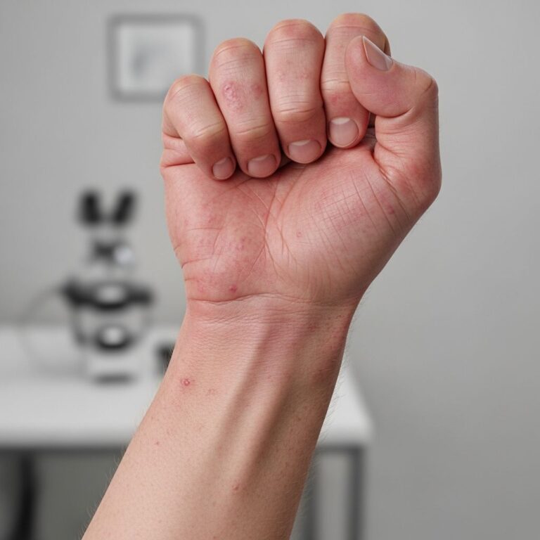
Stretch marks, medically known as striae distensae, are fine linear lines that appear on the skin resulting from rapid growth or over-stretching of tissue beneath the skin surface. These marks develop when the dermis—the layer of skin containing collagen and elastin fibers—tears due to rapid skin expansion. Stretch marks are among the most common benign cutaneous lesions and frequently cause cosmetic and psychological concern in affected individuals.
Stretch marks typically develop in areas of the body subject to rapid stretching, including the abdomen, buttocks, thighs, breasts, back, armpits, and groin. They are usually several centimeters long and measure 1–10 mm in width, though those caused by corticosteroid use or Cushing syndrome are often larger and wider and may involve other regions, including the face.
Causes and Risk Factors
Stretch marks develop due to a combination of mechanical stress and hormonal influences that disrupt the dermal connective tissue. Understanding the underlying causes is essential for both prevention and treatment strategies.
Primary Causes
- Pregnancy: One of the most common causes, with striae frequently developing on the abdomen, breasts, and thighs during gestation.
- Rapid weight changes: Both weight gain and weight loss can trigger stretch mark formation due to sudden skin expansion or contraction.
- Puberty: Growth spurts during adolescence frequently result in stretch marks, particularly in males and females experiencing rapid physical development.
- Muscle hypertrophy: Rapid muscle growth from intensive weight training can cause stretch marks.
- Corticosteroid use: Both topical and systemic corticosteroid exposure can impair fibroblast function and reduce collagen synthesis, predisposing individuals to stretch marks.
- Cushing syndrome: This endocrine disorder causes elevated corticosteroid levels, leading to stretch mark development often characterized by larger, wider marks.
- Genetic disorders: Conditions such as Marfan syndrome and Ehlers-Danlos syndrome increase susceptibility due to inherent connective tissue abnormalities.
Pathophysiological Mechanisms
The development of stretch marks involves several interconnected biological processes. Rapid skin expansion leads to structural damage in the dermis, while elastases released by mast cells and macrophages contribute to the breakdown of elastic tissue. Elevated levels of corticosteroids or genetic predisposition can further impair fibroblast function and reduce collagen synthesis, exacerbating tissue damage.
Types of Stretch Marks
Stretch marks evolve through distinct stages, classified based on their clinical appearance and histological characteristics. Understanding these differences is crucial for determining appropriate treatment approaches.
Striae Rubrae (Red or Purple Stretch Marks)
Striae rubrae represent the early stage of stretch mark development. An early sign is when an area of skin becomes flattened and thin with a pink color, which may occasionally be itchy. Soon after, reddish or purplish slightly swollen lines develop perpendicular to the direction of skin tension. These early lesions exhibit increased vascularity and are characterized by inflammatory changes in the dermis.
Histopathological features of striae rubrae include inflammatory infiltrates, dilated capillaries, and preserved dermal architecture with disrupted collagen and elastin fibers. The red or purple appearance results from increased blood vessel visibility through the thinned, inflamed skin layer.
Striae Albae (White or Flesh-Colored Stretch Marks)
Over months to years, striae rubrae gradually fade into hypopigmented, atrophic, and wrinkled scars known as striae albae. These mature stretch marks resemble atrophic scar tissue and appear whitish or flesh-colored and much less conspicuous than their earlier red or purple counterparts.
Histopathology of striae albae reveals epidermal atrophy, loss of rete ridges, reduced vascularity, and densely packed, thin, scar-like horizontal collagen bundles. Electron microscopy studies have demonstrated mast cell degranulation, macrophage activation, and elastolysis in the mid-dermis. Skin pigmentation is significantly lower in stretch marks compared to adjacent skin.
Clinical Presentation and Diagnosis
Stretch marks present as linear marks that typically run perpendicular to the direction of skin tension and gradually fade over time. The lesions are easily identifiable by their characteristic appearance, and diagnosis is typically clinical without requiring specialized testing.
Dermatological examination may reveal:
- Linear, slightly raised pink to violaceous marks in early stages
- Gradual transition to hypopigmented, atrophic scars in later stages
- Wrinkled appearance and altered skin texture in mature stretch marks
- Skin biopsy showing disrupted collagen and elastin fibers (when histological confirmation is needed)
Stretch marks can occasionally be confused with linear focal elastosis, which has increased elastic fibers on histopathology, though the clinical presentation and distribution patterns typically distinguish between these conditions.
Clinical Significance and Complications
Stretch marks are usually only a cosmetic problem and do not pose significant health risks. However, in rare cases where they are extensive, they may ulcerate or tear easily in an accident. The primary concern for most individuals is the psychological and cosmetic impact of visible stretch marks.
In adolescents, stretch marks become less visible over time and generally require no treatment as the marks naturally fade with age and skin remodeling. However, for individuals seeking to minimize their appearance, various treatment options are available.
Prevention of Stretch Marks
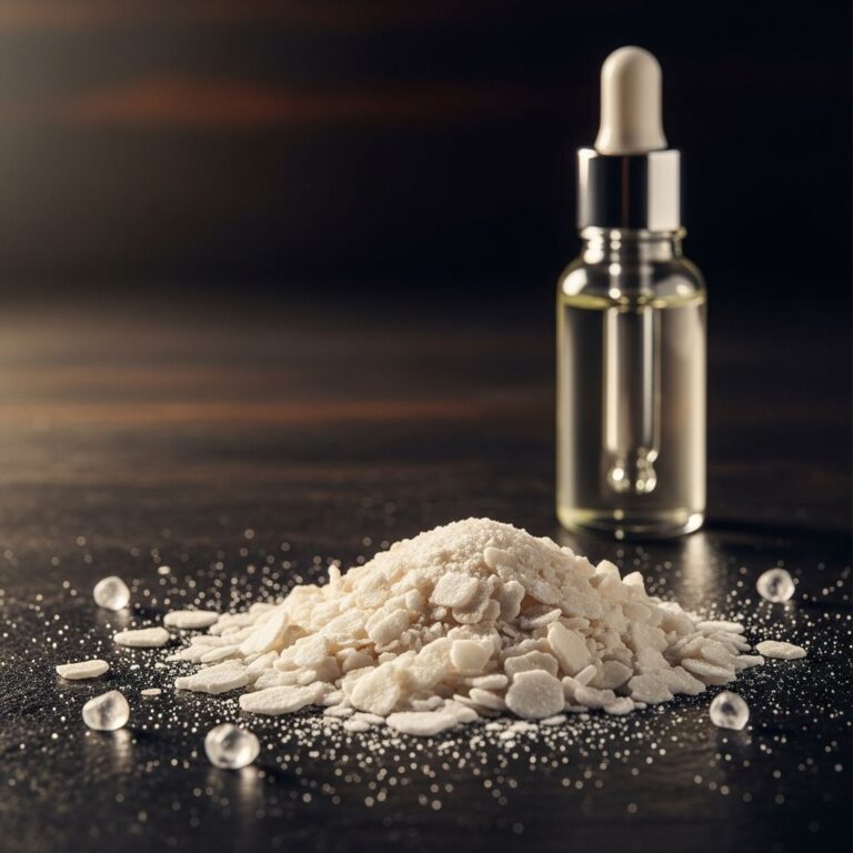
Prevention of stretch marks using topical ointments and oils is debatable, with limited high-quality evidence supporting their effectiveness. Several topical approaches have been investigated for prevention, particularly in pregnant women.
Topical Prevention Strategies
A randomized, double-blind, placebo-controlled study in pregnant women indicates that the severity of stretch marks can be reduced by topical application of emollient and moisturizer containing hydroxyprolisilane C and rose hip oil. However, most clinical trials conducted to date have been of low quality and involved small sample sizes, limiting the conclusiveness of prevention recommendations.
Treatment Options
Multiple therapeutic modalities exist for treating stretch marks, though none can completely eradicate them. The primary goals of treatment differ based on the stage of the stretch mark. For striae rubrae, the focus is on reducing redness, swelling, and irritation. In striae albae, treatment aims to stimulate collagen and elastic fiber production, improve skin hydration, and reduce inflammation.
Non-Laser Treatment Approaches
Topical Treatments
Various topical treatments have been investigated for stretch mark management. In general, early stretch marks (striae rubrae) respond better to treatment than older lesions (striae albae).
Tretinoin Cream: In a prospective randomized open trial, topical 0.05% tretinoin cream was found effective in reducing the severity of early stretch marks. A 12-week regimen is generally required to achieve results, with adverse effects being mild and rare, including local irritation and desquamation. To enhance penetration of topical tretinoid cream, ablative radiofrequency was combined with ultrasound, with pilot trials showing that striae albae improved with this combined approach.
Microdermabrasion
Microdermabrasion was found to be as effective as daily application of topical 0.05% tretinoin cream in reducing the severity of early stretch marks in a prospective randomized open trial. When combined with topical platelet-rich plasma, microdermabrasion proved more effective in reducing stretch mark severity than the single component approach.
Radiofrequency and Electromagnetic Field Treatments
In a pilot study with 16 females, noninvasive multipolar-pulsed electromagnetic field and radiofrequency energy-generating treatment resulted in some improvement in the length and widths of stretch marks.
Microneedling
Microneedling improved both early and late stretch marks in a pilot study carried out among Korean women. This finding is substantiated by a study performed in South Africa and Germany using one to four treatments. Microneedling therapy was more efficient than microdermabrasion with phonophoresis in the treatment of stretch marks.
Laser and Light-Based Treatments
Laser therapy is among the most effective treatment modalities for stretch marks, with different laser types targeting specific pathophysiological features of striae rubrae and striae albae.
Nd:YAG Laser
In a trial with 45 patients, efficacies of two fluences (75 and 100 J/cm²) of long-pulsed Nd:YAG laser on stretch marks were compared using a 5 mm spot size and 15 ms pulse duration. Clinical and histological evaluation performed 3 months after treatment showed that significant improvement in the appearance of striae albae was achieved with 100 J/cm² fluence, whereas striae rubrae showed better improvement with 75 J/cm². Histologically, the dermal content of both collagen and elastin fibers increased following treatment.
Fractional Laser Therapy
Treatment of stretch marks in Asian women with a 1550-nm fractional laser improved stretch marks clinically, with skin elasticity found to be partially normalized. Skin biopsies demonstrated significant increases in epidermal thickness, collagen, and elastic fiber deposition after fractional laser therapy. Adverse effects included mild and transient pain and hyperpigmentation. The Er-doped fractionated 1550-nm laser has been recommended for the treatment of stretch marks in a consensus conference.
Er:YAG Laser
The Er:YAG laser type has been used successfully in patients with stretch marks, with clinically appreciable improvement in striae ranging from 1% to 24%. Three months after the final treatment, patients showed noticeable improvement compared with baseline, though mild post-inflammatory hyperpigmentation was observed in a single patient. The Er:YAG laser has also been used successfully in patients with stretch marks caused by topical corticosteroids. Limited experience exists with the use of fractional Er:YAG laser.
Treatment Mechanisms and Expected Outcomes
In conclusion, laser therapy of early stretch marks (striae rubrae) targets vessels and reduces vascular activity. The treatment of both early and later stretch marks (striae albae) aims to increase collagen production, restore elastin fibers, and restore epidermal thickness. Skin texture improvements also contribute to clinical effects of laser therapy.
The natural course of stretch marks argues for increased vascularity in the early lesions (striae rubrae), supporting the use of vascular lasers for treating red stretch marks. Full clearance of the lesions is very rare and seems to be occasionally obtained only in some isolated areas of recent striae. Therefore, it is vital to start treatment as early as possible, as striae rubrae respond better to therapeutic intervention than the more established striae albae.
Research Gaps and Evidence Limitations
The number of studies with at least 20 patients is limited in the stretch mark literature, and placebo-controlled studies have not been published for many treatment modalities. This represents a significant gap in the evidence base, as most clinical trials have been of low quality with small sample sizes. More rigorous, well-designed clinical trials are needed to definitively establish the efficacy of various treatment approaches for both prevention and management of stretch marks.
Frequently Asked Questions
Q: Are stretch marks dangerous?
A: No, stretch marks are usually only a cosmetic problem and do not pose health risks. In rare cases where they are extensive, they may ulcerate or tear easily in an accident, but this is uncommon.
Q: Can stretch marks be completely removed?
A: No treatment can completely eradicate stretch marks. However, early intervention with appropriate therapies can significantly improve their appearance, particularly when treatment begins during the red or purple stage (striae rubrae).
Q: Do topical treatments prevent stretch marks?
A: Prevention of stretch marks using topical ointments and oils is debatable, with limited high-quality evidence supporting their effectiveness. Some studies suggest that emollients and moisturizers containing specific ingredients may reduce severity, but conclusive evidence is lacking.
Q: Which treatment is most effective for stretch marks?
A: Laser therapy, particularly fractional lasers and Nd:YAG lasers, has shown promising results in treating both early and late stretch marks. Microneedling is also effective. The choice of treatment depends on the type of stretch marks and individual skin characteristics.
Q: Do stretch marks fade on their own?
A: Yes, stretch marks gradually fade over time, especially in adolescents where they often become less visible without treatment. However, they do not completely disappear and may remain visible as pale scars.
Q: What is the best time to treat stretch marks?
A: Early intervention is crucial. Striae rubrae (red or purple stretch marks) respond better to treatment than striae albae (white or flesh-colored marks). It is vital to start treatment as early as possible when the marks first appear.
References
- Management of Stretch Marks with a Focus on Striae Rubrae — Journal of Cosmetic and Aesthetic Surgery. 2019. https://jcasonline.com/management-of-stretch-marks-with-a-focus-on-striae-rubra e/
- Striae Distensae — StatPearls, National Center for Biotechnology Information. 2024. https://www.ncbi.nlm.nih.gov/books/NBK436005/
- Stretch Marks (Striae) — DermNet. 2024. https://dermnetnz.org/topics/stretch-marks-striae
- Topical Management of Striae Distensae (Stretch Marks): Prevention and Therapeutic Options — Journal of Dermatological Venereology. 2016. https://onlinelibrary.wiley.com/doi/10.1111/jdv.13223
Similar Articles


Read full bio of Sneha Tete