Structure Of The Epidermis: Layers, Cells, And Key Functions
Explore the layered architecture of the epidermis, the skin's vital protective barrier against environmental threats.

The
epidermis
is the outermost layer of the skin, serving as a dynamic, semi-permeable barrier that protects the body from environmental hazards, pathogens, and water loss. Composed primarily of keratinocytes, it regenerates continuously through ordered cell division and differentiation, culminating in the flat, anuclear cells of thestratum corneum
.Skin Layers Overview
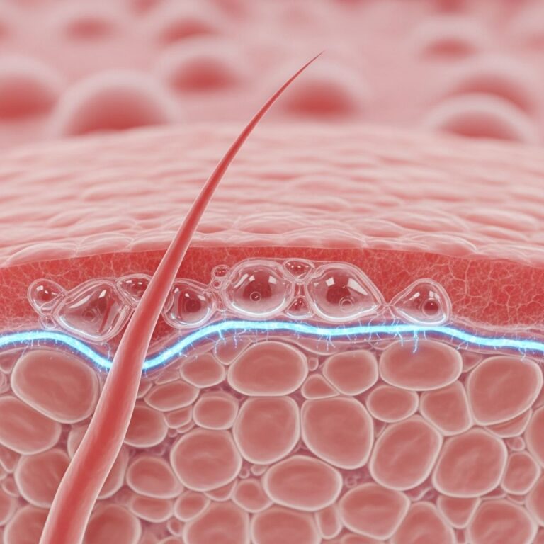
Skin consists of three primary layers: the epidermis, dermis, and subcutis (hypodermis). The epidermis is avascular stratified squamous epithelium, relying on diffusion from the underlying dermis for nutrients. Its thickness varies by body site—thickest on palms and soles (up to 1.5 mm) and thinnest on eyelids (0.05 mm)—adapting to local needs like friction resistance or flexibility.
- Epidermis: Protective barrier with keratinocytes, melanocytes, and immune cells.
- Dermis: Supportive layer with collagen, elastin, vessels, and nerves.
- Subcutis: Fat-rich insulation and cushioning.
Each square centimeter of skin contains approximately 150 cm of blood vessels, 10 hair follicles, 15 sebaceous glands, 100 sweat glands, and over 1,000 nerve endings, highlighting the epidermis’s integration with adnexal structures.
Acid Mantle
The skin surface features an
acid mantle
, a slightly acidic film (pH 4.5–5.5) formed by sebum, sweat, and desquamated corneocytes. This protects against bacterial colonization, maintains barrier integrity, and supports enzymatic activity for desquamation. Disruptions lead to conditions like xerosis or infections.Epidermal Topography
The epidermis exhibits an undulating surface with crisscrossing ridges and valleys, punctuated by invaginations from hair follicles and sweat ducts.
Rete ridges
(epidermal pegs) interlock with dermal papillae, enhancing adhesion. On palms and soles, pronounced ridges form dermatoglyphs (fingerprints).Cell Types in the Epidermis
The epidermis hosts four main cell types, each with specialized functions:
| Cell Type | Location | Function |
|---|---|---|
| Keratinocytes (90%) | All layers | Produce keratin for barrier; regenerate via basal division. |
| Melanocytes | Basal layer | Synthesize melanin in melanosomes to shield from UV; determine skin color. |
| Langerhans cells | Stratum spinosum | Dendritic immune cells; antigen presentation for allergen recognition. |
| Merkel cells | Basal layer | Mechanoreceptors for fine touch; require special stains for visualization. |
Keratinocytes dominate, undergoing keratinization as they migrate upward. Melanocytes transfer melanosomes to keratinocytes via dendrites, providing photoprotection.
Epidermal Layers
The epidermis comprises 4–5 layers (strata) from deep to superficial, with keratinocytes progressing from cuboidal basal cells to flattened corneocytes. Turnover takes ~28 days in young adults, slowing with age.
Basal Cell Layer (Stratum Basale/Germinativum)
The deepest single layer sits atop the basement membrane zone (BMZ). Cuboidal
keratinocytes
divide mitotically, producing daughter cells that migrate upward. It also housesmelanocytes
(1:10 ratio to basal cells),Merkel cells
, and stem cells for renewal. Hemidesmosomes anchor it to the BMZ.- Melanocytes produce melanin, protecting nuclei from UV.
- Merkel cells associate with nerve endings for tactile sensation.
Squamous Cell Layer (Stratum Spinosum/Prickle Cell Layer)
Comprising 8–10 layers of polyhedral keratinocytes connected by
desmosomes
(spinous bridges visible in shrinkage artifacts).Langerhans cells
reside here, aiding immunity. Keratin synthesis begins, and glycolipids are released for waterproofing. Inflammatory cells infiltrate during responses.Desmosomes provide tensile strength, preventing shear.
Granular Cell Layer (Stratum Granulosum)
3–5 layers of flattened keratinocytes containing
keratohyalin granules
(filaggrin precursor) and lamellated bodies (lipid-rich). Odland bodies release lipids forming the mortar of the barrier. Nuclei degrade; cells apoptose.This transition marks the start of the cornified envelope.
Stratum Lucidum
A thin, translucent layer in high-friction areas (palms, soles). Eleidin-filled keratinocytes appear clear under microscopy, enhancing durability.
Horny Cell Layer (Stratum Corneum)
The outermost 15–30 layers of anuclear
corneocytes
embedded in lipid matrix. Fully keratinized, it provides mechanical protection, prevents desiccation, and hosts the acid mantle. Desquamation renews the surface.| Layer | Key Features | Thickness (approx.) |
|---|---|---|
| Stratum corneum | Anuclear corneocytes, lipid mortar | 10–40 μm |
| Stratum granulosum | Granules, lamellated bodies | 5–10 μm |
| Stratum spinosum | Desmosomes, Langerhans cells | 100–200 μm |
| Stratum basale | Mitosis, melanocytes | 10–20 μm |
Basement Membrane Zone (BMZ)
The BMZ anchors epidermis to dermis, facilitating nutrient exchange and mechanical stability. Composed of:
- Basal lamina: Lamina lucida (bullous pemphigoid antigen), lamina densa (type IV collagen).
- Sublamina densa: Anchoring fibrils (type VII collagen) linking to dermal collagen.
Autoimmune diseases like epidermolysis bullosa target BMZ components.
Adnexal Structures
Epidermal invaginations form:
- Eccrine glands: Sweat-producing for thermoregulation.
- Apocrine glands: Scent-related, in axillae/genitals.
- Hair follicles: Cyclic units with sebaceous glands.
- Sebaceous glands: Lipid secretion.
Functions of the Epidermis
Beyond barrier, it enables:
- Photoprotection: Melanin absorbs UV.
- Immunity: Langerhans cells process antigens.
- Sensation: Merkel cells.
- Regeneration: Basal proliferation.
Clinical Relevance
Epidermal alterations underlie psoriasis (hyperproliferation), eczema (barrier defects), and skin cancers (e.g., basal/squamous cell carcinoma from keratinocytes).
Frequently Asked Questions (FAQs)
What is the main function of the stratum corneum?
The stratum corneum acts as the primary physical and chemical barrier, preventing water loss and pathogen entry via keratinized corneocytes and lipids.
How does the epidermis regenerate?
Keratinocytes divide in the basal layer, differentiate upward, and shed from the surface in ~28 days.
What role do melanocytes play?
Melanocytes produce melanin to protect against UV radiation and confer skin pigmentation.
Why is the epidermis thicker on palms and soles?
Enhanced stratum corneum and stratum lucidum provide friction resistance.
What happens if the basement membrane is damaged?
Blistering diseases like pemphigoid occur due to poor adhesion.
References
- Principles of dermatological practice. Structure of the epidermis — DermNet NZ. 2023. https://dermnetnz.org/cme/principles/structure-of-the-epidermis
- Structure of the dermis and subcutis — DermNet NZ. 2023. https://dermnetnz.org/cme/principles/structure-of-the-dermis-and-subcutis
- The structure of normal skin — DermNet NZ. 2023. https://dermnetnz.org/topics/the-structure-of-normal-skin
- Layers of the Skin — Medicine LibreTexts (based on University of Michigan Medical School resources). 2022. https://med.libretexts.org/Bookshelves/Anatomy_and_Physiology/Human_Anatomy_(Lange_et_al.)/04:_Integumentary_System/4.02:_Layers_of_the_Skin
- Anatomy, Skin (Sudoriferous Gland) — StatPearls Publishing LLC (NCBI Bookshelf). 2025. https://www.ncbi.nlm.nih.gov/books/NBK513244/
Similar Articles


Read full bio of medha deb