Subclavian Artery: Anatomy, Function and Health
Understanding the subclavian artery: Its role in blood circulation to arms, head, and neck.

The subclavian artery is a major blood vessel that plays a critical role in delivering oxygen-rich blood to the upper extremities, head, and neck. Located just beneath the clavicle (collarbone), these paired arteries represent essential components of your cardiovascular system. Understanding the anatomy and function of the subclavian artery is important for recognizing potential health concerns and understanding various medical conditions that may affect this vital vessel.
Understanding the Subclavian Artery
The subclavian arteries lie just below the clavicles, providing blood supply to the bilateral upper extremities with contributions to the head and neck. These arteries form part of the main arterial distribution system that branches from the aorta, the body’s largest blood vessel. The subclavian arteries are responsible for channeling nutrient-rich, oxygenated blood to multiple regions of your upper body, ensuring that muscles, tissues, and organs receive the necessary oxygen and nutrients to function properly.
Each side of the body has its own subclavian artery, but they originate from different sources. The right subclavian artery derives from the brachiocephalic trunk, while the left subclavian artery originates directly from the aortic arch. This anatomical difference has important clinical implications, as it can affect how certain conditions develop and progress on each side of the body.
Anatomical Location and Course
The subclavian arteries course laterally between the anterior and middle scalene muscles, traveling from the medial aspect near the thorax toward the lateral shoulder region. The distal limit of the subclavian artery is the lateral border of the first rib, where it becomes the axillary artery. This transition point marks the boundary between two distinct arterial segments that work together to supply the arm and shoulder region.
Located deep within the thorax and shoulder region, the subclavian artery runs alongside important neural structures and venous pathways. The artery forms an interconnected highway that helps fuel the cellular processes used by the neck and upper extremity muscle groups, the brain, and thyroid gland. This proximity to other vital structures means that any disease or abnormality of the subclavian artery can potentially affect these neighboring systems.
Branches of the Subclavian Artery
The subclavian artery gives rise to several important branches that supply different regions of the upper body and head:
– Internal thoracic artery – Supplies the anterior chest wall and breast tissue- Vertebral artery – One of two major arteries responsible for supplying blood to the brain and spinal cord, accounting for approximately 20% of all blood flow to the brain- Costocervical trunk – Supplies the neck and upper back regions- Thyrocervical trunk – Provides blood to the thyroid gland and surrounding neck structures- Dorsal scapular artery – Supplies the scapular region and shoulder muscles
Each of these branches plays a specific role in maintaining adequate blood flow to critical structures. The vertebral arteries are particularly important, as they merge with the internal carotid arteries to form the Circle of Willis, a critical network of vessels that ensures the brain receives continuous blood supply even if one of the major arteries becomes blocked.
Subdivisions of the Subclavian Artery
Given the long path of the artery, the subclavian artery subdivides into three distinct parts, each with different anatomical relationships and branches:
First Part of the Subclavian Artery
The first part initiates as the root of the subclavian artery and ends at the medial edge of the scalene muscles. This area is responsible for feeding the Circle of Willis (providing blood to the brain), thyroid tissue, and breast tissue. The vertebral artery originates from this first segment.
Second Part of the Subclavian Artery
The second part starts at the medial edge of the scalenes and extends to the lateral edge of the scalenes. This middle segment passes directly between the anterior and middle scalene muscles and feeds the costocervical trunk, which supplies various structures in the neck and thorax.
Third Part of the Subclavian Artery
The third part initiates at the lateral scalene muscle and extends to the lateral tip of the first rib, at which point the artery becomes the axillary artery. This distal segment feeds the upper extremity through its continuation as the axillary artery, which eventually branches into the brachial, radial, and ulnar arteries.
Surrounding Structures and Relationships
Multiple aspects of the nervous system travel alongside or near the subclavian arteries. These include the sympathetic trunk, the vagus nerve, parts of the brachial plexus, the phrenic nerve, and the right recurrent laryngeal nerve. The close proximity of these neural structures explains why certain subclavian artery conditions can produce neurological symptoms such as voice changes or arm weakness.
Alongside these neuronal pathways, the arteries are linked closely to venous pathways such as the internal jugular veins and vertebral veins. The subclavian artery runs parallel to the subclavian vein, which carries oxygen-poor blood back toward the heart. These paired vessels work together as a coordinated system, with the artery delivering fresh blood and the vein removing deoxygenated blood for recirculation through the lungs.
Embryological Development
The development of the subclavian arteries is unique to each anatomical side. During fetal development, the left subclavian artery develops differently than the right, which has important implications for understanding anatomical variations:
Left Subclavian Artery Development
The fully developed left subclavian artery arises from only the seventh intersegmental artery during embryological development. This relatively straightforward origin explains why the left subclavian artery typically has a more predictable anatomy compared to its right-sided counterpart.
Right Subclavian Artery Development
The right side is unique as the three parts of the subclavian develop from three different embryological vessels. The first part of the right subclavian develops from the right fourth aortic arch, part of one of the six paired great embryologic vessels. The middle and distal segments develop from different embryological sources, creating a more complex developmental pattern than the left side. This embryological complexity explains why anatomical variations are more common on the right side.
Anatomical Variations
The most common variant of the subclavian artery is the aberrant subclavian, which means the artery root comes off at a location different from the typical anatomy. The majority of these cases are benign and asymptomatic, requiring no treatment or intervention. However, in some cases, the variant may cause symptomatology.
The most common aberrant variant occurs when the right subclavian artery roots directly from the aortic arch as the last branch of the arch. In this configuration, it travels posteriorly and may wrap around the esophagus, leading to a vascular ring that can compress the esophagus. This compression may induce dysphagia lusoria, a condition characterized by difficulty swallowing, particularly with solid foods. Individuals with this variant may experience symptoms ranging from mild swallowing difficulty to more severe esophageal compression requiring intervention.
Clinical Significance and Common Conditions
Given the extensive supply territory of the subclavian arteries and their proximity to vital structures, numerous conditions can occur either through congenital defects, years of pathologic change, or idiopathic causes.
Subclavian Artery Stenosis
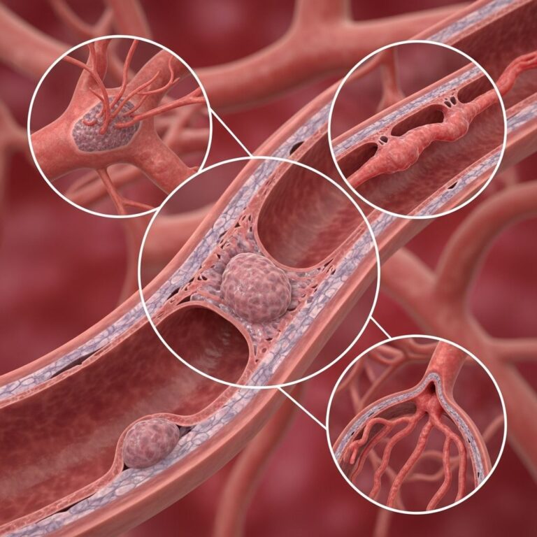
Subclavian artery stenosis refers to the narrowing of this blood vessel, usually because of plaque buildup. This condition makes it difficult to get the blood flow needed for proper upper extremity function. Many people do not experience symptoms because other arteries can compensate by sending blood to the arm and making up for the slowdown in the subclavian artery. However, symptomatic stenosis represents a warning sign that plaque buildup is occurring in your arteries.
Subclavian Steal Syndrome
Subclavian steal syndrome (SSS) is a condition that sends blood to the arm instead of the brain because of narrowing in the subclavian artery. As this blockage worsens, blood moves through the subclavian artery with less pressure. Stronger pressure from nearby arteries makes blood flow backward in a vertebral artery that branches off the subclavian artery, diverting blood intended for the brain to supply the arm instead. This phenomenon can cause symptoms related to reduced brain blood flow, arm symptoms, or both. Most people with subclavian steal syndrome do not experience symptoms, but when present, symptoms warrant medical evaluation and management of underlying risk factors.
Aberrant Subclavian Artery
An aberrant subclavian artery is one that does not arise from the normal location and therefore has an altered pathway in the thorax. Although symptoms are usually benign, in some cases the artery may cause esophageal narrowing due to a vascular ring compressing the esophagus, or voice changes from pressure placed on the recurrent laryngeal nerve.
Blood Flow and Circulation
As major branches within the aortic arterial supply line, the subclavian arteries provide vital flow for much of the head, neck, and upper extremities. The distribution of blood through the subclavian system represents a carefully balanced system that ensures adequate oxygen delivery to all structures supplied by these vessels. When one pathway becomes compromised, collateral circulation from alternative blood vessels may partially compensate, explaining why some individuals with significant subclavian artery disease may have minimal or no symptoms.
Frequently Asked Questions
Q: What does the subclavian artery supply?
A: The subclavian artery supplies blood to the upper extremities, head, neck, brain (via the vertebral artery), thyroid gland, and breast tissue. Each branch of the subclavian artery delivers oxygen-rich blood to specific regions of the upper body.
Q: Why do the right and left subclavian arteries originate differently?
A: The right subclavian artery derives from the brachiocephalic trunk, while the left subclavian artery originates directly from the aortic arch. This difference in embryological development explains anatomical variations between the two sides.
Q: What happens if the subclavian artery becomes blocked?
A: Depending on the degree of blockage and the body’s ability to develop collateral circulation, symptoms may range from none to significant arm weakness, reduced brain blood flow (in subclavian steal syndrome), or swallowing difficulties. Medical evaluation is necessary to determine appropriate treatment.
Q: What neural structures are near the subclavian artery?
A: The brachial plexus, vagus nerve, phrenic nerve, sympathetic trunk, and right recurrent laryngeal nerve all travel near or alongside the subclavian artery, which explains why subclavian artery pathology can sometimes cause neurological symptoms.
Q: Where exactly is the subclavian artery located?
A: The subclavian artery is located just beneath the clavicle (collarbone), coursing between the anterior and middle scalene muscles before becoming the axillary artery at the level of the first rib.
References
- Anatomy, Thorax, Subclavian Arteries — National Center for Biotechnology Information (NCBI). 2024. https://www.ncbi.nlm.nih.gov/books/NBK539736/
- Subclavian Vein: Location, Anatomy and Function — Cleveland Clinic. 2024. https://my.clevelandclinic.org/health/body/23941-subclavian-vein
- Vertebral Artery: What Is It, Location, Anatomy and Function — Cleveland Clinic. 2024. https://my.clevelandclinic.org/health/body/21689-vertebral-artery
- Subclavian Steal Syndrome (SSS): Symptoms & Causes — Cleveland Clinic. 2024. https://my.clevelandclinic.org/health/diseases/subclavian-steal-syndrome
- Subclavian Artery Stenosis: Symptoms & Treatment — Cleveland Clinic. 2024. https://my.clevelandclinic.org/health/diseases/subclavian-artery-stenosis-and-disease
- Arteries: What They Are, Anatomy & Function — Cleveland Clinic. 2024. https://my.clevelandclinic.org/health/body/22896-arteries
- What Is the Brachiocephalic Artery? — Cleveland Clinic. 2024. https://my.clevelandclinic.org/health/body/24410-brachiocephalic-artery
Similar Articles


Read full bio of Sneha Tete