Understanding Superior Limbic Keratoconjunctivitis
A comprehensive overview of SLK: causes, symptoms, diagnosis, and evidence-based treatment approaches

Superior Limbic Keratoconjunctivitis: Clinical Insights and Management Strategies
Superior limbic keratoconjunctivitis, commonly abbreviated as SLK, represents a distinctive inflammatory condition affecting the ocular surface, primarily involving the superior bulbar conjunctiva and cornea. This condition has emerged as an important diagnostic consideration in contemporary clinical ophthalmology, affecting individuals across various demographic groups and presenting with variable severity levels. Understanding the pathophysiology, recognition of clinical manifestations, and implementation of appropriate management strategies are essential components of comprehensive eye care.
Defining the Condition and Its Epidemiological Context
Superior limbic keratoconjunctivitis is characterized by inflammation localized to the superior limbal region of the eye, the transitional zone between the cornea and sclera at the upper aspect of the eyeball. The condition manifests as chronic or recurrent inflammation affecting both the conjunctival and corneal tissues in this specific anatomical location. While the exact prevalence remains incompletely understood, clinicians recognize SLK as an important subset of ocular surface disease, frequently encountered in both general and specialized ophthalmologic practices.
The condition occurs across all age groups, though certain patterns of presentation suggest variable risk among populations. The chronic or intermittent nature of SLK distinguishes it from acute inflammatory conditions, requiring sustained clinical attention and appropriate long-term management strategies. Recognition of this entity has improved significantly over recent decades as clinical knowledge and diagnostic capabilities have advanced.
Underlying Pathophysiological Mechanisms
The pathophysiology of superior limbic keratoconjunctivitis involves complex interactions between mechanical, inflammatory, and potentially immune-mediated factors. Mechanical trauma to the superior conjunctival tissue represents one proposed mechanism, potentially resulting from repetitive contact with the upper eyelid during blinking or eye movements. This mechanical irritation may trigger an inflammatory cascade, leading to persistent conjunctival and corneal changes.
The superior limbic region’s unique anatomical characteristics contribute to its susceptibility to inflammation. This area experiences constant exposure to environmental stressors and undergoes continuous mechanical stress during normal eye movements. Additionally, the limbal vasculature and innervation patterns in the superior region may render it particularly vulnerable to inflammatory processes.
Some research suggests potential associations between SLK and underlying systemic conditions, including thyroid disorders and connective tissue diseases. The proposed mechanisms involve autoimmune phenomena affecting ocular tissues, though these associations require further investigation. Environmental factors, including exposure to irritants and allergens, may also contribute to disease initiation or exacerbation in susceptible individuals.
Clinical Presentation and Symptomatology
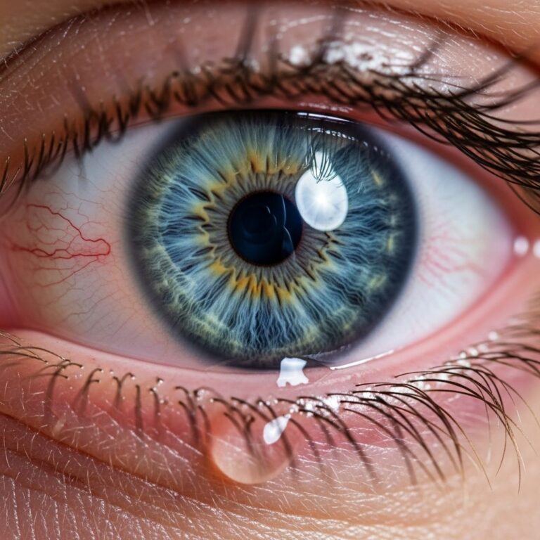
Patients with superior limbic keratoconjunctivitis typically report ocular discomfort characterized by a foreign body sensation, mild irritation, and grittiness affecting the upper aspect of the eye. The symptom severity varies considerably among affected individuals, ranging from minimal discomfort to more pronounced irritation affecting visual comfort and daily functioning. Many patients describe symptoms that fluctuate throughout the day or vary with environmental conditions.
Visual disturbances may accompany the physical discomfort, including transient blurred vision or mild photophobia. Some individuals report discharge, though this typically remains minimal. The chronic nature of symptoms frequently leads patients to seek medical evaluation, as self-directed remedies often prove insufficient for symptom control.
The following characteristic features frequently present in SLK cases:
- Conjunctival injection (redness) in the superior tarsal region
- Mild discharge or mucus production
- Conjunctival papillae formation on the upper tarsal surface
- Superior corneal involvement ranging from punctate epithelial defects to filamentary keratitis
- Limbal hyperemia and thickening
- Cyclical variation in symptom severity
Diagnostic Evaluation and Clinical Assessment
Establishing a diagnosis of superior limbic keratoconjunctivitis relies primarily on comprehensive slit-lamp biomicroscopic examination, which reveals the characteristic anatomical and inflammatory changes. The diagnostic approach emphasizes direct visualization of superior conjunctival and limbal pathology while assessing the extent of corneal involvement.
Clinical examination typically demonstrates the following diagnostic features:
- Superior conjunctival hyperemia and edema
- Papillary reaction on the upper tarsal conjunctiva
- Rose bengal or lissamine green staining of the superior conjunctiva and limbus
- Corneal staining patterns indicating epithelial irregularities or filaments
- Normal intraocular pressure and anterior chamber examination
Additional diagnostic considerations include careful assessment of tear film quality and quantity to exclude concurrent aqueous tear deficiency. Ocular surface staining procedures, employing vital dyes such as lissamine green, contribute to diagnostic confirmation by highlighting areas of conjunctival and corneal epithelial disruption in the superior region.
The diagnostic process must also eliminate alternative diagnoses presenting with similar features, including seasonal or perennial allergic conjunctivitis, vernal keratoconjunctivitis, and dry eye disease. Historical information regarding symptom onset, progression pattern, and relationship to seasons or environmental exposures assists in differential diagnosis. Associated systemic symptoms or medical conditions warrant consideration when present.
Management and Therapeutic Approaches
Treatment strategies for superior limbic keratoconjunctivitis aim to reduce inflammation, promote ocular surface healing, and provide symptomatic relief. The approach typically progresses from conservative measures to more intensive interventions, depending on disease severity and response to initial management.
Conservative and Topical Management: Initial therapeutic approaches emphasize lubrication and topical anti-inflammatory intervention. Artificial tear preparations, particularly those containing mucopolysaccharides or hyaluronic acid, provide symptomatic relief and protect the ocular surface. Lubricating ointments, especially when applied before sleep, reduce overnight drying and may decrease morning symptom severity.
Topical corticosteroid agents represent an important component of management for moderate to severe inflammation. These medications effectively reduce conjunctival and corneal inflammatory changes, though prolonged use requires careful monitoring for steroid-related complications including elevated intraocular pressure and corneal changes. Intermittent corticosteroid use, alternating with corticosteroid-sparing agents, frequently becomes the preferred long-term strategy.
Advanced Topical Therapies: Topical immunosuppressive agents have emerged as valuable corticosteroid-sparing alternatives. Cyclosporine ophthalmic emulsion reduces inflammatory responses through immune modulation without the adverse effects associated with long-term steroid use. This medication proves particularly beneficial for chronic disease management.
Nonsteroidal anti-inflammatory drugs (NSAIDs) in topical formulation may complement other therapies, though extended use carries considerations regarding corneal integrity. These agents effectively manage inflammation and associated discomfort while preserving long-term safety profiles more favorable than prolonged corticosteroid therapy.
Procedural and Surgical Interventions: Refractory cases may benefit from more definitive therapeutic approaches. Cauterization of the superior conjunctiva, performed chemically or thermally, has demonstrated efficacy in reducing inflammation and preventing recurrence. This procedure addresses the pathophysiological basis of disease by modifying the involved tissue, thereby reducing inflammatory potential.
Conjunctival debridement or superficial keratectomy may be considered for advanced cases with significant corneal involvement or scarring. These procedures remove diseased tissue while promoting regeneration of healthier epithelium. Such interventions require careful patient selection and consideration of potential visual outcomes.
Comparative Treatment Options and Evidence Considerations
| Treatment Modality | Mechanism of Action | Typical Duration | Monitoring Considerations |
|---|---|---|---|
| Artificial Tears | Lubrication and surface protection | Ongoing as needed | Minimal; frequency adjustment based on symptoms |
| Topical Corticosteroids | Anti-inflammatory suppression | 2-4 weeks initially, then tapered | Intraocular pressure monitoring; long-term use avoided |
| Cyclosporine | Immune modulation | 8-12 weeks for therapeutic effect | Periodic assessment of response; excellent long-term safety |
| Conjunctival Cauterization | Tissue modification and inflammatory reduction | Single or repeated procedures | Follow-up assessment for recurrence; resolution rates variable |
Long-term Prognosis and Disease Course
The long-term trajectory of superior limbic keratoconjunctivitis varies substantially among affected individuals. Some patients experience resolution with appropriate management, while others develop chronic disease requiring ongoing therapeutic intervention. Understanding this variability helps establish realistic expectations and guides long-term management planning.
Factors influencing prognosis include disease severity at presentation, response to initial therapy, associated systemic conditions, and anatomical variations in limbal tissue. Early diagnosis and prompt initiation of appropriate treatment improve outcomes and reduce the risk of advanced corneal changes or permanent scarring.
Recurrence represents a recognized feature of SLK, with some patients experiencing periodic exacerbations despite successful initial treatment. Maintenance therapy using corticosteroid-sparing agents often becomes necessary to prevent symptom recurrence and progressive tissue damage.
Frequently Asked Questions Regarding Superior Limbic Keratoconjunctivitis
Is superior limbic keratoconjunctivitis contagious? No, SLK is not contagious. It represents an inflammatory condition rather than an infectious process, posing no transmission risk to other individuals.
Can SLK cause permanent vision loss? While SLK typically does not cause permanent vision impairment, advanced cases with significant corneal scarring or epithelial damage may result in mild visual disturbances. Early appropriate treatment minimizes this risk substantially.
What role does thyroid disease play in SLK development? Some evidence suggests associations between thyroid disorders and SLK occurrence, though causative mechanisms remain incompletely understood. Patients with thyroid conditions warrant careful ocular surface assessment.
How long does treatment typically continue? Treatment duration varies considerably. Some patients respond to therapy and remain stable after several months, while others require ongoing management. Long-term corticosteroid-sparing approaches often become the preferred strategy for chronic cases.
Are environmental factors important in SLK management? Yes, environmental modifications including reduced exposure to irritants, appropriate humidity levels, and protective eyewear may complement medical management and improve symptom control.
Clinical Considerations for Contemporary Practice
Superior limbic keratoconjunctivitis remains an important diagnostic entity requiring recognition and appropriate management by contemporary ophthalmologists. The condition’s variable presentation and chronic nature demand sustained clinical attention and individualized therapeutic approaches. Advances in understanding disease mechanisms and availability of multiple therapeutic options have substantially improved management outcomes.
Integration of mechanical, inflammatory, and potentially immune-mediated perspectives on disease pathogenesis informs rational therapeutic selection. Patient education regarding disease chronicity, treatment expectations, and the importance of consistent care enhances adherence and improves long-term outcomes. Multidisciplinary approaches incorporating both medical and procedural interventions optimize results for complex or refractory disease presentations.
References
- American Academy of Ophthalmology Cornea/External Disease Panel — American Academy of Ophthalmology. 2023. https://www.aao.org
- Ocular Surface Disease Study (OSDS) Guidelines — The Ocular Surface Society. 2023. https://www.theocularsurfacesociety.org
- PubMed Central Ophthalmology Literature Database — National Library of Medicine, U.S. National Institutes of Health. 2024. https://www.ncbi.nlm.nih.gov/pmc
- Wills Eye Hospital Manual of Ophthalmology — Wolters Kluwer Health. 2024. https://www.wolterskluwer.com
- Journal of Ophthalmology Peer-Reviewed Research Articles — Hindawi Publishing Corporation. 2024. https://www.hindawi.com/journals/joph
Similar Articles


Read full bio of Sneha Tete