SVC Obstruction Syndrome: Causes, Symptoms, And Treatment
Understand the causes, symptoms, diagnosis, and treatments for superior vena cava obstruction, a critical condition often linked to cancer.

The superior vena cava (SVC) serves as a vital conduit, transporting deoxygenated blood from the head, neck, arms, and upper chest directly to the heart’s right atrium. When this major vein faces narrowing or complete blockage, it results in SVC obstruction syndrome, a condition that demands prompt medical attention. This syndrome arises from external compression, internal thrombosis, or direct invasion, leading to impaired venous return and a cascade of distressing symptoms.
Understanding the Anatomy and Pathophysiology
The SVC, the body’s second-largest vein, originates at the confluence of the brachiocephalic veins and descends through the mediastinum, just posterior to the sternum. Its thin, compliant walls make it susceptible to compression by nearby structures such as lymph nodes, tumors, or mediastinal masses. In SVC syndrome, venous pressure upstream rises dramatically, causing collateral circulation to develop over time, though acute cases overwhelm these compensatory mechanisms.
Pathophysiologically, the blockage triggers venous hypertension in the upper body, manifesting as edema and engorged veins. Acute obstructions develop rapidly, often within days, while chronic ones progress over weeks, allowing partial adaptation via alternative pathways like the azygos vein.
Primary Causes and Risk Factors
Malignant causes dominate, accounting for the majority of cases. Lung cancer, particularly small cell and non-small cell types, is the leading culprit, with tumors directly invading or compressing the SVC. Lymphoma, including non-Hodgkin types, and metastases from breast, testicular, or gastrointestinal cancers also contribute significantly by enlarging mediastinal lymph nodes.
- Lung malignancies: Responsible for up to 60-80% of cases due to proximity in the chest.
- Lymphoproliferative disorders: Such as Hodgkin and non-Hodgkin lymphoma affecting central nodes.
- Metastatic disease: From distant primaries spreading to thoracic lymphatics.
Non-malignant etiologies, though less common today, include thrombosis from indwelling catheters (e.g., central venous lines for chemotherapy), fibrosing mediastinitis from histoplasmosis, or aortic aneurysms. Benign causes have declined with improved catheter care protocols.
Risk factors amplify susceptibility: smoking history for lung cancer, prior mediastinal radiation, or chronic central venous access. Patients with advanced thoracic malignancies face the highest risk.
Recognizing the Warning Signs
Symptoms stem from venous congestion and vary by obstruction acuity. Gradual onset allows symptom acclimation, whereas sudden blockage presents as an emergency.
| Symptom Category | Common Manifestations | Frequency |
|---|---|---|
| Respiratory | Shortness of breath, cough, hoarseness | Most common (up to 60%) |
| Facial/Neck | Swelling (face, neck), headache, dizziness | 50-60% |
| Upper Limb/Chest | Arm edema, visible chest/neck veins | 30-50% |
| Neurological/Visual | Visual changes, confusion (severe cases) | 10-20% |
Classic signs include facial plethora (redness), periorbital edema worsening when supine, and prominent neck veins. Severe cases risk laryngeal edema, airway compromise, or cerebral edema. In children, rapid progression heightens airway obstruction risk, making it life-threatening.
Diagnostic Approaches
Diagnosis combines clinical suspicion with imaging. Initial evaluation includes chest X-ray for widening mediastinum or pleural effusions. Contrast-enhanced CT scans with venography provide definitive visualization of SVC narrowing, collateral vessels, and underlying pathology.
- CT chest with contrast: Gold standard, detects 95% of cases.
- MRI: Useful for contraindications to contrast or detailed soft tissue assessment.
- Venography: Invasive, reserved for procedural planning.
- Biopsy: Tissue diagnosis via bronchoscopy, mediastinoscopy, or EBUS for malignancies.
Elevated serum tumor markers or cytopenias may hint at lymphoma. Multidisciplinary input from oncology, radiology, and interventional specialists is essential.
Management and Treatment Options
Treatment prioritizes symptom relief, SVC patency restoration, and addressing the underlying cause. Supportive measures precede definitive therapy.
Supportive Care
- Head elevation (30-45 degrees) to reduce venous pressure.
- Supplemental oxygen for dyspnea.
- Corticosteroids (e.g., dexamethasone) to decrease peritumoral edema.
- Diuretics cautiously; loop diuretics for significant edema.
- Anticoagulation if thrombosis present, avoiding in hemorrhagic risks.
Definitive Interventions
For malignant SVCO, systemic therapy targets the primary disease. Chemotherapy and radiation achieve response in 70-90% of small cell lung cancer cases, though relief may lag 1-2 weeks.
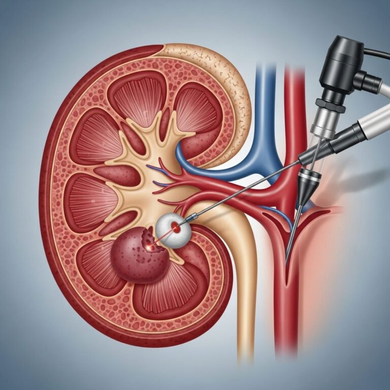
SVC Stenting: Minimally invasive gold standard for rapid palliation, success rates exceeding 90%. Performed by interventional radiologists under fluoroscopy, stents (self-expanding metal) are deployed via femoral or jugular access under local anesthesia or sedation. Complications (5-10%) include migration, thrombosis, or rupture, managed with adjunctive therapies.
Surgery (bypass grafting) is rare, reserved for benign, refractory cases due to high morbidity.
Prognosis and Long-Term Outlook
Outcomes hinge on etiology and treatment timeliness. Malignant SVCO carries guarded prognosis tied to cancer stage; median survival 6-12 months post-diagnosis with therapy. Benign cases often resolve fully. Stenting confers immediate symptom relief in 80-95%, with patency 6-24 months. Follow-up monitors recurrence via imaging and symptom review.
Quality of life improves markedly with intervention; palliative care integrates for advanced disease, focusing on dyspnea control and psychosocial support.
Prevention Strategies
While not always preventable, risks mitigate via smoking cessation, vigilant catheter management (prompt removal post-use), and early thoracic malignancy screening in high-risk groups.
Frequently Asked Questions (FAQs)
What triggers SVC obstruction most frequently?
Lung cancer predominates, followed by lymphoma and metastases.
Is SVC syndrome immediately life-threatening?
Potentially yes, if airway or cerebral compromise occurs; urgent care is advised.
How effective is stenting for relief?
Highly, with >90% symptom improvement shortly post-procedure.
Can non-cancer causes lead to this?
Yes, including clots from catheters or infections like histoplasmosis.
What lifestyle changes help post-treatment?
Elevate head, avoid straining, monitor for recurrence.
References
- Superior vena cava obstruction – CIRSE — Cardiovascular and Interventional Radiological Society of Europe. 2023. https://www.cirse.org/patients/general-information/medical-conditions/superior-vena-cava-obstruction/
- Superior vena cava obstruction in palliative care – Marie Curie — Marie Curie. 2024. https://www.mariecurie.org.uk/professionals/palliative-care-knowledge-zone/superior-vena-cava-obstruction
- Superior vena cava obstruction (SVCO) — Macmillan Cancer Support. 2024. https://www.macmillan.org.uk/cancer-information-and-support/impacts-of-cancer/superior-vena-cava-obstruction
- Superior Vena Cava Syndrome (SVCS) — Cleveland Clinic. 2023-10-27. https://my.clevelandclinic.org/health/diseases/23304-superior-vena-cava-syndrome
- Superior vena cava syndrome — Canadian Cancer Society. 2024. https://cancer.ca/en/treatments/side-effects/superior-vena-cava-syndrome
- SVC obstruction – Health Encyclopedia — HealthFinder Florida. 2023. https://quality.healthfinder.fl.gov/health-encyclopedia/HIE/1/001097
Similar Articles


Read full bio of medha deb