Systemic Amyloidosis: Early Signs, Diagnosis, Treatment Guide
Uncommon disorder of misfolded proteins causing organ dysfunction, with key skin signs in AL type alerting to systemic disease.

Systemic amyloidosis is an uncommon disorder in which misfolded proteins become resistant to normal processes of removal by the body. Their accumulation in bodily organs leads to abnormal organ function. The heart, liver, kidneys, nerves, lungs, and bowel may be affected, and in only one type of systemic amyloidosis (AL type) is the skin significantly affected and indeed may be the clue to discovering underlying internal organ disease.
What is systemic amyloidosis?
Systemic amyloidosis is subclassified according to the chemical composition of the misfolded protein. There are two components to the amyloid:
- A protein (e.g., light chains in AL amyloidosis)
- Serum amyloid P (common to all types)
Systemic amyloidosis is further classified into types, including AL (immunoglobulin light chain), AA (secondary to chronic inflammation), ATTR (transthyretin-related), and others like dialysis-related or hereditary forms. Chronic inflammation due to autoinflammatory conditions, autoimmune disease, and infections may induce AA systemic amyloid (see section below). This does not produce skin signs or symptoms but may be identified histologically in subcutaneous fat. Overall worldwide, AA amyloidosis outnumbers all other forms due to high prevalence of infectious diseases in developing countries.
Primary systemic amyloidosis (AL amyloidosis) is a plasma-cell dyscrasia of unknown cause. Immunoglobulin light chains, or fragments of light chains, produced by plasma-cell clones form extracellular amyloid fibrils. Amyloid deposition can occur in any organ in the body, causing features such as congestive cardiac failure, renal failure and hepatosplenomegaly, as well as skin lesions. Both sexes are affected by this disease, with onset most commonly from the sixth decade. Although recent advances in therapy are encouraging, the prognosis for primary amyloidosis remains poor.
Who gets systemic amyloidosis?
Systemic amyloidosis affects individuals across ages but is more common in older adults, particularly over 50 years for AL type. AL amyloidosis typically presents in the sixth decade or later, with equal incidence in males and females. AA amyloidosis is linked to chronic inflammatory conditions like rheumatoid arthritis, infections (e.g., tuberculosis in endemic areas), or familial Mediterranean fever. Risk factors include plasma cell dyscrasias for AL, chronic infections or autoinflammatory diseases for AA, and genetic mutations for hereditary forms.
Clinical features
Extracellular deposition of insoluble amyloid protein in affected organs as well as the skin and its adnexae causes tissue destruction leading to dysfunction. However, the precise mechanism of how amyloid fibril is formed and sequestrated into tissues has not been well understood yet. Cutaneous manifestations will vary depending on the site of amyloid deposition and the degree of local tissue destruction; it occurs in 30–40% of patients with primary systemic AL amyloidosis.
Usual features include:
- Purpura: Periorbital (‘raccoon eyes’), post-pinching purpura (roof-thrust sign), easy bruising
- Waxy papules and nodules: Eyelids, face, neck, limbs; may coalesce into plaques
- Plaques: Waxy, indurated; mimic scleroderma
- Bullae: Tense, haemorrhagic; legs
- Alopecia: Non-scarring
- Nail dystrophy
- Macroglossia: Firm, indolent enlargement (20% AL cases)
Direct dermal infiltration can produce subcutaneous nodules and plaques. Classically described as smooth, waxy, yellowish lesions, these are uncommon and more often appear hemorrhagic. They are generally found on flexor surfaces, the face, and the buccal mucosa. Cutaneous nodules can be demonstrated to follow the path of blood vessels within the skin. Direct skin infiltration with amyloid can produce the appearance of scleroderma on the face, hands, and feet. Rarely, infiltrative nodular lesions may coalesce to form gross distortion and enlargement or tumefaction.
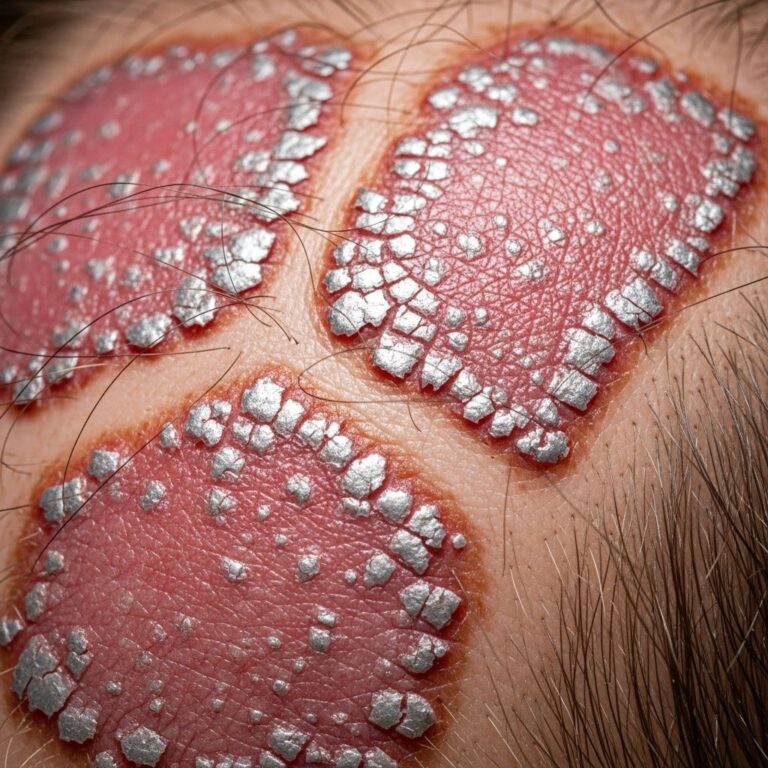
The most common skin lesions in primary systemic amyloidosis, i.e., petechiae, purpura, and ecchymosis, are those related to intracutaneous hemorrhage due to infiltration of blood vessel wall by amyloid. In some cases, unusual presentations occur, such as multiple skin-colored nodules (2-5 cm) over scalp, trunk, and extremities, coalescing into plaques resembling cutis verticis gyrata, or noduloulcerative tongue lesions mimicking squamous cell carcinoma.
Complications
The complications are variable depending on the site and extent of involvement. Common organ-specific issues include:
- Cardiac: Heart failure, arrhythmias, sudden death
- Renal: Nephrotic syndrome, proteinuria, renal failure
- Neurologic: Peripheral neuropathy, autonomic dysfunction
- Hepatic: Hepatomegaly, elevated liver enzymes
- Gastrointestinal: Malabsorption, bleeding, motility issues
Congestive cardiac failure is a particularly poor prognostic factor. Amyloid deposits up to the deep dermis can result in nodular lesions, and bone marrow involvement indicates plasma cell dyscrasias.
Diagnosis
Should the dermatologist be presented with early skin stigmata, there may be a valuable opportunity for early diagnosis should the disease be included appropriately in a working differential on initial consultation. Intervention may then be initiated before end-organ failure, allowing the possibility of a wider range of treatment options and greater opportunity to delay the development of disease. The greatest prognostic factor is the stage of disease at the time of initial treatment, demonstrating that early diagnosis is of paramount importance.
Skin biopsy characteristically shows diffuse amyloid deposition in the form of nodules and plaques. There may be amyloid infiltration of blood-vessel walls, around individual fat cells, and in pilosebaceous units. Amyloid deposits have also been identified in the nailfold and bed of dystrophic nails. These changes may also often be seen in biopsies of clinically normal skin. Distinction between systemic and localized forms of amyloid on skin biopsy does not usually present serious difficulty.
Key diagnostic steps:
- Congored staining of biopsy: Apple-green birefringence under polarized light
- Subcutaneous abdominal fat aspiration (preferred non-invasive for systemic detection)
- Bone marrow biopsy for plasma cell dyscrasia in AL type
- Serum/urine immunofixation, free light chains
- Organ function tests (echo, renal biopsy if needed)
Herein, hematoxylin, and eosin followed by Congo red staining confirmed the diagnosis. In this case, the amyloid deposits up to the deep dermis resulted in nodular lesions. Bone marrow biopsy is done to find out plasma cell dyscrasias. Subcutaneous abdominal fat aspiration is the preferred method for detecting systemic amyloidosis.
Differential diagnosis
The differential diagnosis will depend on presenting features, and include:
| Feature | Differential Diagnoses |
|---|---|
| Periorbital purpura | Trauma, anticoagulation, dermatomyositis |
| Waxy papules/nodules | Lipoid proteinosis, mucinosis, xanthomas |
| Macroglossia | Acromegaly, hypothyroidism |
| Scleroderma-like plaques | Scleromyxoedema, nephrogenic systemic fibrosis |
| Pinch purpura | Ehlers-Danlos syndrome, actinic purpura |
Skin involvement can occur as primary cutaneous amyloidosis or as a part of systemic amyloidosis. In localized cutaneous disease, the amyloidogenic protein is produced at the site of deposition.
Treatment
The prognosis for AL amyloidosis is poor, with death usually resulting from cardiac or renal failure. Sudden death from cardiac arrhythmias also occurs. The mean survival from diagnosis is approximately 15 months. The average time from identification of systemic AL amyloidosis to death is several months if left untreated. Some treatments, including stem cell transplantation and chemotherapy, have achieved 10-year survival periods.
Treatment is aimed at decreasing amyloid production and deposition and promoting lysis of deposits. Response to treatment may be complete, partial, or negative. Current treatment options include:
- Chemotherapy: Oral melphalan with/without prednisone
- Stem cell transplant: Autologous stem-cell harvest and transfer with high-dose melphalan
- Proteasome inhibitors: Bortezomib-based regimens
- Immunomodulators: Lenalidomide, daratumumab
- Supportive: Organ-specific (diuretics for heart failure, dialysis for renal)
Other agents like dimethyl sulfoxide or colchicine have been described, although with less success. For AA amyloidosis, treat underlying inflammation (e.g., anti-TNF for rheumatoid arthritis).
Prognosis
Cardiac and renal amyloidosis are poor prognostic features. Early diagnosis significantly improves outcomes, as the stage at treatment initiation is the key factor. With modern therapies like stem cell transplant, long-term survival (up to 10 years) is possible in select cases.
Systemic AA amyloidosis
Chronic inflammation of the skin can rarely induce systemic AA amyloidosis, and chronic pustular psoriasis and psoriatic arthritis are perhaps the best established causes. AA amyloidosis arises from serum amyloid A protein elevated in chronic inflammation. Unlike AL, skin signs are absent, but subcutaneous fat may show deposits. Management focuses on controlling the underlying disease to halt progression.
Frequently Asked Questions
What causes systemic amyloidosis?
Misfolded proteins (e.g., light chains in AL type from plasma cells, serum amyloid A in AA from inflammation) deposit extracellularly.
Can skin signs be the first clue?
Yes, in 30-40% of AL cases; purpura, waxy papules prompt biopsy and systemic workup.
Is amyloidosis curable?
Not curative, but early treatment can achieve remission and prolong survival; stem cell transplant offers best outcomes in eligible patients.
How is it diagnosed?
Biopsy with Congo red (apple-green birefringence), fat aspirate, serum free light chains, bone marrow exam.
What is the prognosis?
Poor if advanced (months survival); better with early intervention (years possible).
References
- Primary, systemic amyloidosis and the dermatologist: Where classic skin lesions are the presenting features of disease — eScholarship, University of California. 2005. https://escholarship.org/uc/item/4kp7580w
- Systemic Amyloidosis: Cutaneous features — DermNet NZ. Recent update (authoritative dermatology resource). https://dermnetnz.org/topics/systemic-amyloidosis
- Primary Systemic Amyloidosis with Unusual Dermatological Manifestations — NIH, PMC. 2016-04-01. https://pmc.ncbi.nlm.nih.gov/articles/PMC4817453/
Similar Articles


Read full bio of Sneha Tete