Targetoid Haemosiderotic Haemangioma Pathology
Detailed pathology of rare benign angiomas with targetoid appearance and hobnail endothelial cells.

Targetoid haemosiderotic haemangiomas, also known as hobnail haemangioma or targetoid haemangioma, are rare benign vascular lesions characterized by a distinctive targetoid clinical appearance and unique histopathological features. These angiomas typically present as small erythematous or violaceous papules or macules surrounded by a pale or ecchymotic ring, most commonly on the trunk and extremities of young to middle-aged adults.
Clinical features
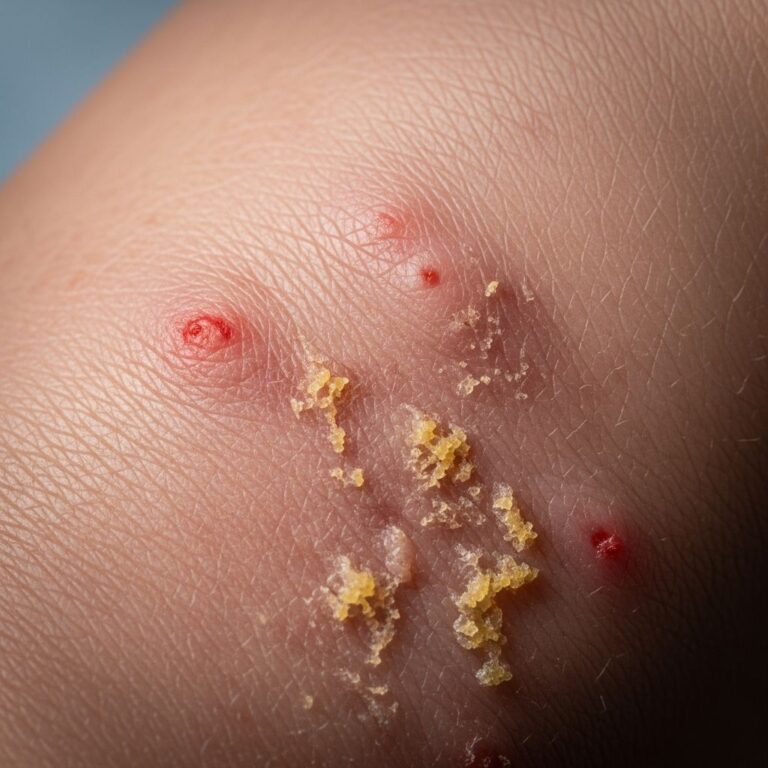
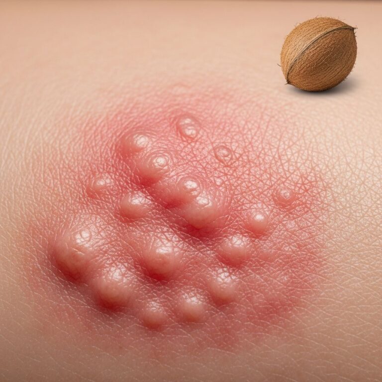
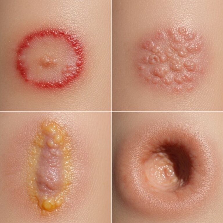
Clinically, targetoid haemosiderotic haemangioma manifests as a solitary lesion, usually less than 1 cm in diameter, with a central purple-brown papule encircled by a thin pale area and an outer ecchymotic (bruise-like) halo, creating the classic targetoid morphology. The lesion often arises de novo or following minor trauma, with the peripheral ring expanding centrifugally before resolving, leaving a persistent central papule. Sites of predilection include the trunk and extremities, though facial and other locations have been reported. Dermoscopy reveals red or reddish-blue lacunae, haemorrhagic crusts as black macules, and sometimes arborizing vessels in atypical cases. The condition is benign, with lesions remaining stable or undergoing spontaneous regression of the halo over time.
Histopathology
Histopathological examination reveals a biphasic growth pattern that is diagnostic for targetoid haemosiderotic haemangioma. In the superficial dermis, ectatic vascular spaces lined by prominent hobnail endothelial cells—characterized by their protruding, hobnail-like nuclei—are prominent, often forming small haemangiomatous nodules around adnexal structures like sweat glands. These superficial vessels show dilated lumina with extravasated erythrocytes and occasional fibrin thrombi.
Deeper in the dermis, the lesion transitions to narrow, angulated, slit-like vascular channels that dissect between collagen bundles, resembling lymphatic vessels. Endothelial cells here are less epithelioid, with intraluminal papillary projections. Stromal changes include haemosiderin deposition (evident on Perl’s iron stain), fibrosis, and a mixed inflammatory infiltrate with lymphocytes. In later stages, the lesion may show a collapsed appearance with increased fibrosis and prominent siderophages. Recent immunohistochemical evidence supports a lymphatic malformation origin rather than a true neoplasm, with positive staining for lymphatic markers like D2-40 alongside vascular markers.
Histopathology images
- Low-power view: Symmetrical lesion with biphasic pattern—superficial dilated vessels and deep collagen-dissecting channels.
- Hobnail endothelial cells: Prominent in superficial ectatic vessels, protruding into lumina.
- Deep dermal component: Slit-like spaces dissecting collagen with haemosiderin deposits.
Immunohistochemistry
The vascular nature is confirmed by positivity for endothelial markers CD31 and CD34, which highlight both superficial and deep components. Lymphatic markers such as D2-40 (podoplanin) are often positive, supporting the lymphatic malformation hypothesis. HHV-8 is negative, distinguishing it from Kaposi sarcoma. Ki-67 proliferative index is low, indicating benignity.
Differential diagnosis
Targetoid haemosiderotic haemangioma enters several clinical and histological differentials due to its evolving appearance and vascular features.
Clinical differentials
- Melanocytic lesions: Targetoid appearance mimics halo naevus or melanoma.
- Inflammatory conditions: Erythema multiforme, insect bite reaction.
- Other vascular lesions: Infantile haemangioma, tufted angioma, dermatofibroma.
Histological differentials
| Feature | Targetoid Haemosiderotic Haemangioma | Angiosarcoma | Kaposi Sarcoma |
|---|---|---|---|
| Endothelial cells | Hobnail, mild atypia | Marked atypia, pleomorphism | Slit-like, no hobnail |
| Architecture | Biphasic, superficial dermal | Infiltrative, deep | Collagen dissection, spindle cells |
| Haemosiderin | Prominent | Rare | Variable |
| IHC | CD31+, D2-40+, HHV-8- | CD31+, high Ki-67 | HHV-8+ |
Angiosarcoma: Distinguished by significant nuclear atypia, deep infiltration, and high mitotic activity; THH lacks these aggressive features. Kaposi sarcoma: Shares deep slit-like spaces but lacks hobnail cells and is HHV-8 positive. Other considerations include progressive lymphangioma and epithelioid haemangioma.
Management
As a benign lesion, intervention is reserved for diagnostic confirmation or cosmetic concerns. Simple excision is curative and provides definitive histology. Laser therapy (e.g., pulsed dye laser) is an effective non-invasive option for persistent lesions. Observation is appropriate for asymptomatic cases, given the low risk of progression.
Frequently Asked Questions (FAQs)
Is targetoid haemosiderotic haemangioma cancerous?
No, it is a benign vascular malformation with no malignant potential.
What causes the targetoid appearance?
Minor trauma leads to haemorrhage and haemosiderin deposition, forming the characteristic halo.
Does it resolve on its own?
The peripheral ring often fades spontaneously, but the central papule may persist.
How is it diagnosed?
Biopsy with histopathology is gold standard; dermoscopy aids clinical suspicion.
Is treatment always necessary?
No, excision or laser is optional for cosmesis or confirmation.
References
- Targetoid haemosiderotic haemangioma: dermoscopic monitoring of three cases — Sahin MT et al. Journal of the European Academy of Dermatology and Venereology. 2005-10-01. https://pubmed.ncbi.nlm.nih.gov/16197386/
- Targetoid haemosiderotic haemangioma pathology — DermNet NZ (Assoc Prof Patrick Emanuel). 2014. https://dermnetnz.org/topics/targetoid-haemosiderotic-haemangioma-pathology
- Targetoid hemosiderotic hemangioma — Wikipedia (sourced from primary literature). N/A. https://en.wikipedia.org/wiki/Targetoid_hemosiderotic_hemangioma
- Cutaneous Vascular Anomalies: Targetoid Hemosiderotic Hemangioma — Perris Dermatology. N/A. https://perridermatology.com/dr-perris-blog/cutaneous-vascular-anomalies-targetoid-hemosiderotic-hemangioma/
- Arborizing vessels in a targetoid hemosiderotic hemangioma — Enei ML et al. Dermatology Practical & Conceptual. 2017. https://dpcj.org/index.php/dpc/article/view/dermatol-pract-concept-articleid-dp0701a08
- Targetoid hemosiderotic hemangioma — VisualDx. N/A. https://android7.visualdx.com/visualdx/diagnosis/targetoid+hemosiderotic+hemangioma?diagnosisId=56175&moduleId=101
Similar Articles


Read full bio of Sneha Tete