Telangiectasia: 5 Proven Treatments, Causes, And Types
Understanding visible small dilated blood vessels: causes, types, diagnosis, and effective treatment options.

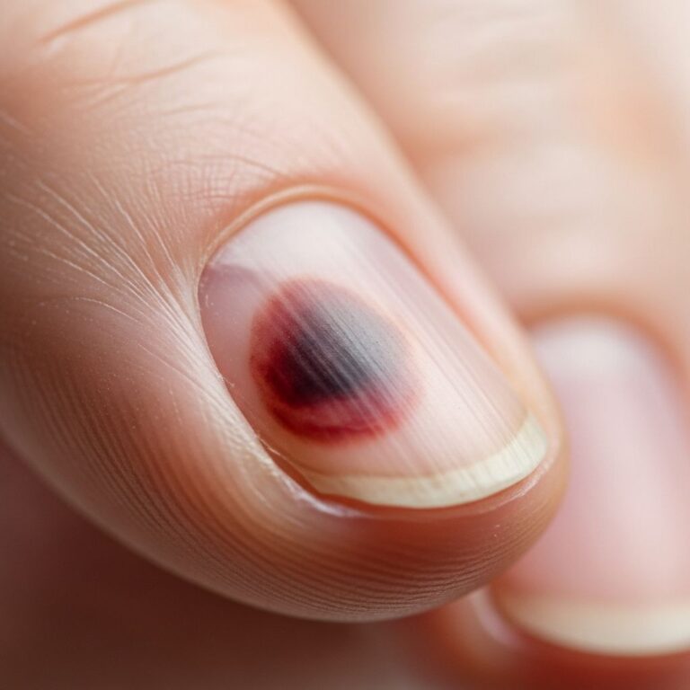
Telangiectasia refers to small, widened blood vessels that are visible near the surface of the skin, often appearing as fine red, blue, or purple lines or webs. These are commonly known as spider veins or broken capillaries and are typically harmless, though they can cause cosmetic concerns or signal underlying health issues.
What is telangiectasia?
Telangiectasias are permanently dilated small blood vessels, including capillaries, venules, or arterioles, located close to the skin’s surface. Unlike normal vessels, they lose tone and become visibly enlarged, forming linear patterns, clusters, or spider-like shapes. They most commonly appear on the face (especially cheeks, nose, and chin), legs, chest, back, and arms but can occur anywhere on the body. While usually asymptomatic, they may occasionally bleed, itch, or ache, particularly on the legs.
In medical terms, telangiectasia derives from Greek roots meaning ‘end vessel expansion,’ accurately describing the condition where these superficial vessels fail to constrict properly. They differ from varicose veins, which involve larger, deeper veins, but can coexist with venous insufficiency.
Who gets telangiectasia?
Telangiectasia affects individuals across all ages, genders, and ethnicities, but certain groups are more prone. Fair-skinned people, women (due to hormonal influences), and those over 30 are at higher risk. Prevalence increases with age, sun exposure, and family history.
- General population: Common in 10-20% of adults, often on facial areas from chronic sun damage.
- Pregnant women: Hormonal changes and increased blood volume lead to temporary leg and facial telangiectasias.
- Those with rosacea: Up to 75% develop facial telangiectasias as a hallmark feature.
- Autoimmune conditions: Frequent in scleroderma (especially CREST syndrome), lupus, and dermatomyositis.
- Genetic disorders: Hereditary hemorrhagic telangiectasia (HHT) affects 1 in 5,000-10,000 people, causing recurrent nosebleeds and mucocutaneous lesions.
- Rare syndromes: Ataxia-telangiectasia (A-T) impacts 1 in 40,000-100,000, with ocular and skin telangiectasias alongside neurological symptoms.
What causes telangiectasia?
Telangiectasia results from vessel wall weakening or dilation due to various triggers. Primary causes are genetic, while secondary factors exacerbate the condition.
Primary causes
- Genetic predisposition: Inherited traits affect vessel elasticity, as seen in HHT from mutations causing arteriovenous malformations.
Secondary causes
- Sun and wind exposure: Chronic UV damage degrades collagen around vessels, common on cheeks and nose.
- Hormonal factors: Pregnancy, oral contraceptives, or hormone replacement therapy dilate vessels.
- Medications: Prolonged corticosteroids (oral/topical) or vasodilators weaken vessel walls.
- Alcohol excess: Chronic use damages skin and vessels.
- Skin trauma: Surgery, injury, acne, or rosacea scars lead to localized dilation.
- Systemic diseases: Liver disease, venous insufficiency, or autoimmune disorders like scleroderma impair vascular repair.
In scleroderma, telangiectasias may reflect compensatory increased blood flow to poorly perfused tissues. In A-T, ATM gene mutations disrupt DNA repair, leading to progressive vascular changes.
What are the clinical features of telangiectasia?
Telangiectasias present as asymptomatic red, purple, or blue lines (0.1-1 mm wide) or webs. Facial ones are linear or arborizing; leg types often radiate from a central vessel (spider telangiectasia).
| Type | Appearance | Common Sites |
|---|---|---|
| Linear | Straight red lines | Cheeks, nose |
| Arborizing | Branching, tree-like | Legs, thighs |
| Spider | Central arteriole with radiating venules | Face, proximal legs |
| Matted | Cluster forming pink/red patch | Legs (associated with veins) |
Symptoms are rare but include itching, burning, or bleeding if traumatized. In HHT, telangiectasias on lips, tongue, and fingers bleed easily, causing anemia. A-T features zig-zag conjunctival vessels and sun-sensitive skin changes.
Diagnosis
Diagnosis is clinical, based on visible lesions and history. Dermoscopy reveals hairpin, comma, or glomerular loops.
- History: Onset, family history, triggers (sun, hormones), associated symptoms (nosebleeds in HHT).
- Examination: Blanching on pressure confirms vascular nature.
- Investigations: For systemic causes: ANA for autoimmune, genetic testing for HHT/A-T, endoscopy for GI bleeding. Leg ultrasound rules out venous reflux.
How is telangiectasia treated?
Treatment is optional for cosmetics but essential if bleeding or symptomatic. Options target vessel coagulation or sclerosis.
Conservative measures
- Sun protection (SPF 50+), avoid irritants, leg elevation/compression stockings.
Procedural treatments
| Method | Best For | Pros/Cons |
|---|---|---|
| Laser (PDL, IPL, Nd:YAG) | Facial, small vessels | Non-invasive, 70-90% clearance; multiple sessions, pigmentation risk |
| Sclerotherapy | Leg telangiectasias | Effective for networks; injections, bruising, recurrence |
| Intense pulsed light (IPL) | Face, rosacea-associated | Broad coverage; less precise than laser |
| Micropulse 1064 nm laser | Delicate areas | Gentle, minimal downtime |
| Electrodesiccation | Small lesions | Simple; scarring possible |
For HHT, treatments include laser for epistaxis, embolization for AVMs. A-T management is supportive, focusing on ataxia and infections. Recurrence is common; maintenance sessions advised.
What is the outcome for telangiectasia?
Benign telangiectasias persist without progression but may multiply with triggers. Cosmetic treatments offer long-term improvement (1-5 years), though new ones form. In diseases like HHT, monitoring prevents complications like anemia or strokes. Early intervention improves quality of life.
Prevention
- Use broad-spectrum sunscreen daily.
- Avoid extreme temperatures, trauma.
- Manage weight, exercise for leg circulation.
- Treat rosacea promptly.
- Genetic counseling for hereditary forms.
Frequently Asked Questions
Are telangiectasias dangerous?
Usually not; they are cosmetic. Rarely, they bleed or indicate systemic disease like HHT.
Do telangiectasias go away on their own?
No, they are permanent but can fade post-treatment.
Can creams treat spider veins?
Topicals may camouflage but don’t eliminate; procedures are needed.
Is telangiectasia hereditary?
Often has genetic component, especially in HHT or familial cases.
How many laser sessions for facial telangiectasia?
Typically 2-4, spaced 4-6 weeks apart.
References
- Hereditary hemorrhagic telangiectasia – Symptoms and causes — Mayo Clinic. 2023-10-15. https://www.mayoclinic.org/diseases-conditions/hht/symptoms-causes/syc-20351135
- Ataxia-telangiectasia: Symptoms, Causes and Outlook — Cleveland Clinic. 2024-05-20. https://my.clevelandclinic.org/health/diseases/23415-ataxia-telangiectasia
- Telangiectasia: Causes, diagnosis, and treatment — Medical News Today. 2023-11-10. https://www.medicalnewstoday.com/articles/312845
- Telangiectasia: Causes, Diagnosis, Prevention & Treatment — Hospital for Special Surgery (HSS). 2024-02-28. https://www.hss.edu/health-library/conditions-and-treatments/telangiectasia-and-autoimmune-disease
- Telangiectasia — MedlinePlus (U.S. National Library of Medicine). 2024-01-05. https://medlineplus.gov/ency/article/003284.htm
Similar Articles


Read full bio of medha deb