Thrombosis: Causes, Symptoms, and Treatment
Understanding blood clots: Learn about thrombosis symptoms, causes, diagnosis, and effective treatment options.

Understanding Thrombosis: A Comprehensive Guide
Thrombosis is a serious medical condition characterized by the formation of a blood clot, or thrombus, within the blood vessels or chambers of the heart. This abnormal clot formation can severely restrict or completely block blood flow, potentially leading to life-threatening complications such as heart attacks, strokes, and pulmonary embolism. Understanding thrombosis is critical because it represents a significant global health burden, being the underlying cause of approximately 1 in 4 deaths worldwide. Early recognition and treatment of thrombosis can substantially improve outcomes and prevent devastating complications.
What Is Thrombosis?
Thrombosis occurs when blood clots form abnormally within blood vessels or the heart. Unlike normal clotting, which is essential for stopping bleeding from injuries, thrombotic clots form without an apparent need and can obstruct blood flow. When a blood clot develops, it may remain stationary at its site of origin or break loose and travel through the bloodstream to lodge in another location. This mobility of clots makes thrombosis particularly dangerous, as a clot that travels to critical organs like the lungs, brain, or heart can cause immediate life-threatening emergencies. The condition requires prompt medical attention and ongoing management to prevent recurrence and complications.
The formation of blood clots involves a delicate balance between coagulation factors, blood cells, platelets, plasma proteins, and the integrity of blood vessel linings. When this balance is disrupted, thrombosis can develop. Understanding the underlying mechanisms helps healthcare providers predict risk and develop appropriate prevention and treatment strategies.
Types of Thrombosis
Thrombosis is classified into two primary categories based on the type of blood vessel affected: arterial thrombosis and venous thrombosis. Each type presents distinct clinical characteristics, risk factors, and treatment approaches.
Arterial Thrombosis
Arterial thrombosis occurs when blood clots form within arteries, the vessels responsible for carrying oxygen-rich blood from the heart to body tissues. When an arterial clot forms, it reduces oxygen delivery to organs and tissues, potentially causing ischemia and tissue death (necrosis). Most commonly, arterial thrombosis affects the coronary arteries of the heart or cerebral arteries in the brain. Arterial thrombosis is frequently caused by atherosclerosis, a condition characterized by plaque buildup in artery walls. Patients experiencing acute arterial thrombosis in the coronary arteries often report crushing left-sided chest pain with radiation to the arm or jaw, representing a medical emergency requiring immediate intervention.
Venous Thrombosis
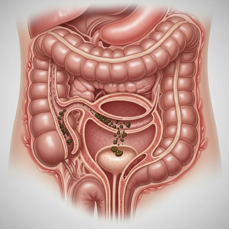
Venous thrombosis refers to blood clot formation within veins, the vessels that return blood to the heart. This category includes deep vein thrombosis (DVT), which occurs in deep veins usually located in the legs, thighs, pelvis, or arms. Venous thrombosis is a common precursor to pulmonary embolism (PE), a life-threatening condition where a blood clot travels to the lungs and blocks pulmonary blood vessels. Unlike arterial thrombosis, venous thrombosis develops more gradually and may produce less dramatic initial symptoms, making it potentially underdiagnosed.
Causes and Risk Factors
Thrombosis results from an imbalance in the body’s natural blood clotting mechanisms. Historically, medical science has recognized three primary factors that predispose individuals to thrombosis, collectively known as Virchow’s triad: damage to blood vessel linings, reduced blood flow, and hypercoagulability (increased tendency of blood to clot).
Primary Causes
Blood clots form when the inner lining of blood vessels (endothelium) becomes damaged or when blood flow slows significantly. Damage to vessel linings triggers inflammatory responses that promote platelet activation and adhesion molecule expression, initiating the coagulation cascade. Reduced blood flow allows blood components to accumulate and stick together abnormally, creating clots. These mechanisms can be triggered by various conditions and lifestyle factors.
Medical Conditions Associated with Thrombosis
Certain medical conditions substantially increase thrombosis risk. Individuals with atrial fibrillation, a heart rhythm disorder, face elevated risk due to irregular blood flow in the heart chambers. Diabetes impairs endothelial function and increases blood clotting tendency. Cancer patients experience hypercoagulability because tumor cells express procoagulant proteins and tissue factors that activate the clotting cascade. Coronary artery disease (CAD) predisposes patients to arterial thrombosis through atherosclerotic plaque disruption. Additional conditions increasing risk include heart failure, recent surgery or hospitalization, autoimmune disorders, and inherited clotting disorders.
Additional Risk Factors
Beyond medical conditions, numerous factors contribute to thrombosis development. Obesity increases blood viscosity and promotes inflammation, both prothrombotic processes. Immobility following bone fractures, surgery, or prolonged bed rest significantly elevates risk through blood stasis. Certain medications, including oral contraceptives and hormone replacement therapy, contain compounds that enhance clotting tendency. Smoking damages endothelial cells and promotes atherosclerosis. Advanced age, pregnancy, and recent trauma also increase thrombosis risk. Understanding these multifactorial causes is essential for developing comprehensive prevention strategies tailored to individual risk profiles.
Symptoms and Clinical Presentations
Thrombosis symptoms vary considerably depending on clot location, size, and the organ systems affected. Some individuals may experience no noticeable symptoms, particularly with small clots in vessels with good collateral circulation. Others present with dramatic acute symptoms requiring emergency care. Location-specific symptoms provide important diagnostic clues.
Symptoms by Location
Pulmonary Embolism (Lungs)
When clots lodge in lung arteries, patients typically experience sudden shortness of breath, chest pain aggravated by breathing, rapid heart rate, and sometimes fainting. Severe pulmonary embolism can cause acute respiratory distress and hemodynamic collapse.
Stroke or Transient Ischemic Attack (Brain)
Cerebral thrombosis produces acute neurological symptoms including sudden unilateral weakness or paralysis, facial drooping, speech difficulty, confusion, vision changes, severe headache, difficulty ambulating, and sensory disturbances. These symptoms demand immediate emergency evaluation.
Heart Attack (Coronary Arteries)
Coronary thrombosis causes crushing chest pain or heaviness, often radiating to the left arm, jaw, or back. Associated symptoms include shortness of breath, nausea, diaphoresis, and overwhelming anxiety.
Abdominal Pain (Mesenteric Vessels)
Thrombosis affecting abdominal blood vessels causes severe abdominal pain, often out of proportion to physical examination findings, along with nausea and vomiting.
Limb Symptoms (Arterial or Venous)
Leg or arm thrombosis presents with localized pain, swelling, warmth, redness, or skin discoloration. Arterial thrombosis may cause coolness, pallor, and muscle pain with minimal exertion. Deep vein thrombosis frequently causes unilateral leg swelling and calf tenderness.
Diagnosis of Thrombosis
Accurate diagnosis of thrombosis requires a combination of clinical evaluation, laboratory testing, and imaging studies. Healthcare providers consider patient history, physical examination findings, and risk factors when determining diagnostic approach.
Diagnostic Methods
Imaging Studies: Ultrasound is the primary diagnostic tool for deep vein thrombosis, providing real-time visualization of clots within veins. Computed tomography pulmonary angiography (CTPA) is the gold standard for diagnosing pulmonary embolism. CT angiography and magnetic resonance angiography visualize arterial vasculature and reveal clots, aneurysms, dissections, and anatomic abnormalities. Cardiac catheterization is typically performed when myocardial infarction is suspected.
Laboratory Tests: D-dimer testing, which measures fibrin degradation products, helps exclude thromboembolism in low-risk patients. Prothrombin time (PT) and activated partial thromboplastin time (aPTT) assess coagulation function. Complete blood counts evaluate platelet levels and hematocrit. Specific testing for inherited thrombophilias or antiphospholipid syndrome may be indicated based on clinical presentation.
Clinical Scoring: Risk stratification tools help clinicians estimate thrombosis probability and guide diagnostic decision-making. The Wells score and Geneva score assist in assessing pulmonary embolism probability.
Complications of Untreated Thrombosis
Failure to recognize and treat thrombosis promptly can result in serious complications. Arterial thrombosis causing myocardial infarction can lead to cardiogenic shock, arrhythmias, and sudden cardiac death. Cerebral thrombosis produces stroke with potential permanent neurological disability, including paralysis, speech impairment, and cognitive changes. Pulmonary embolism can cause acute respiratory failure, hemodynamic instability, and death. Venous thrombosis may result in post-thrombotic syndrome, characterized by chronic limb pain, swelling, and skin changes. Recurrent thrombosis is a significant risk for untreated or inadequately managed patients.
Treatment Approaches
Thrombosis treatment depends on clot location, size, chronicity, symptoms, and underlying provoking factors. Treatment strategies generally progress through three stages: initial acute phase management, chronic phase anticoagulation, and extended secondary prevention.
Acute Phase Management
Initial treatment aims to prevent clot propagation and embolization while restoring adequate blood flow. Anticoagulant medications including heparin or low-molecular-weight heparin are administered intravenously or subcutaneously to inhibit further clot formation. In emergency situations involving massive pulmonary embolism or acute myocardial infarction, thrombolytic therapy (clot-busting drugs) may be employed to actively dissolve existing clots, though this approach carries increased bleeding risk.
Chronic Phase Management
Following the acute phase, patients transition to warfarin, a vitamin K antagonist, or newer direct oral anticoagulants (DOACs) for ongoing anticoagulation. These medications maintain therapeutic anticoagulation to prevent recurrent thrombosis while allowing natural clot dissolution and recanalization of affected vessels. Duration of anticoagulation varies based on provoked versus unprovoked thrombosis and individual risk factors.
Interventional Procedures
Some patients benefit from catheter-directed thrombolysis or mechanical thrombectomy, procedures in which interventional radiologists or cardiologists mechanically remove or dissolve clots. These approaches are considered when anticoagulation alone is insufficient or contraindicated. Inferior vena cava filters may be placed in select patients with contraindications to anticoagulation to prevent pulmonary embolism.
Prevention Strategies
Secondary prevention focuses on identifying and managing underlying provoking factors. Patients with transient provocation (surgery, immobility) may receive limited-duration anticoagulation, while those with persistent risk factors or unprovoked thrombosis typically require extended anticoagulation. Lifestyle modifications including regular physical activity, smoking cessation, compression stockings for venous disease, and weight management support pharmacologic treatment.
Prevention of Thrombosis
Thrombosis prevention is critical, as it is often preventable through risk factor modification and, when indicated, pharmacologic prophylaxis. Patients at high risk for thrombosis benefit from comprehensive risk assessment and individualized prevention strategies.
General prevention measures include maintaining healthy body weight, regular physical activity, smoking cessation, and adequate hydration. Patients undergoing surgery or facing prolonged immobility should receive venous thromboembolism prophylaxis with anticoagulants or mechanical devices. Individuals with atrial fibrillation, prior thrombosis, or inherited thrombophilias may benefit from long-term anticoagulation. Compression therapy helps prevent venous stasis in at-risk individuals.
Living with Thrombosis
Patients with thrombosis require ongoing medical management and lifestyle adjustments. Regular monitoring with laboratory tests ensures appropriate anticoagulation levels. Patient education about recognizing warning signs of recurrent thrombosis or bleeding complications is essential. Patients should understand medication adherence importance, maintain consistent anticoagulation, and report symptoms promptly. Support groups and patient resources provide valuable education and peer support for individuals managing thrombotic conditions.
Frequently Asked Questions
Q: Can thrombosis be cured?
A: Thrombosis itself often resolves with appropriate anticoagulation therapy, allowing natural clot dissolution. However, individuals with unprovoked thrombosis may require lifelong anticoagulation to prevent recurrence. Treatment success depends on early intervention and compliance with prescribed medications.
Q: How long does thrombosis treatment take?
A: Initial acute phase treatment lasts several days to weeks. Anticoagulation duration varies from 3 months for provoked thrombosis to indefinite therapy for unprovoked cases or those with persistent risk factors. Your healthcare provider determines the appropriate duration based on individual circumstances.
Q: What foods interact with blood thinners?
A: Foods high in vitamin K, including leafy greens like kale, spinach, and broccoli, interact with warfarin. Maintaining consistent vitamin K intake rather than complete avoidance is recommended. Direct oral anticoagulants have fewer food interactions. Discuss dietary concerns with your healthcare provider.
Q: Can I exercise with thrombosis?
A: Physical activity is generally beneficial and encouraged during thrombosis treatment and recovery. Begin gradually under medical supervision, and avoid strenuous activity immediately after diagnosis. Walking, swimming, and other low-impact exercises improve circulation and reduce thrombosis risk.
Q: What are warning signs of serious complications?
A: Seek emergency care for sudden severe chest pain, shortness of breath, sudden severe headache, facial drooping, speech difficulty, sudden weakness, severe abdominal pain, or heavy bleeding. These symptoms may indicate life-threatening complications requiring immediate evaluation.
Q: Is thrombosis hereditary?
A: Some thrombosis forms result from inherited blood clotting disorders, such as Factor V Leiden or prothrombin gene mutations. If multiple family members have experienced thrombosis, genetic testing may be indicated. Discuss family history with your healthcare provider.
References
- About Venous Thromboembolism (Blood Clots) — Centers for Disease Control and Prevention (CDC). 2024. https://www.cdc.gov/blood-clots/about/index.html
- Thrombosis — StatPearls, National Center for Biotechnology Information (NCBI). 2024. https://www.ncbi.nlm.nih.gov/books/NBK538430/
- Deep Vein Thrombosis (DVT) — Symptoms & Causes — Mayo Clinic. 2024. https://www.mayoclinic.org/diseases-conditions/deep-vein-thrombosis/symptoms-causes/syc-20352557
- Thrombosis: Symptoms & Treatment — Cleveland Clinic. 2024. https://my.clevelandclinic.org/health/diseases/22242-thrombosis
- Deep Vein Thrombosis (DVT) — Northwestern Medicine. 2024. https://www.nm.org/conditions-and-care-areas/vein-center/deep-vein-thrombosis
Similar Articles


Read full bio of Sneha Tete